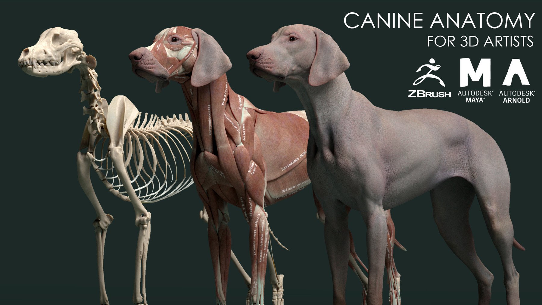
ArtStation Canine Anatomy Model Resources
Labeled anatomy of the head and skull of the dog on CT imaging (bones of cranium, brain, face, paranasal sinus, muscles of head) This module of vet-Anatomy presents an atlas of the anatomy of the head of the dog on a CT. Images are available in 3 different planes (transverse, sagittal and dorsal), with two kind of contrast (bone and soft tissues).

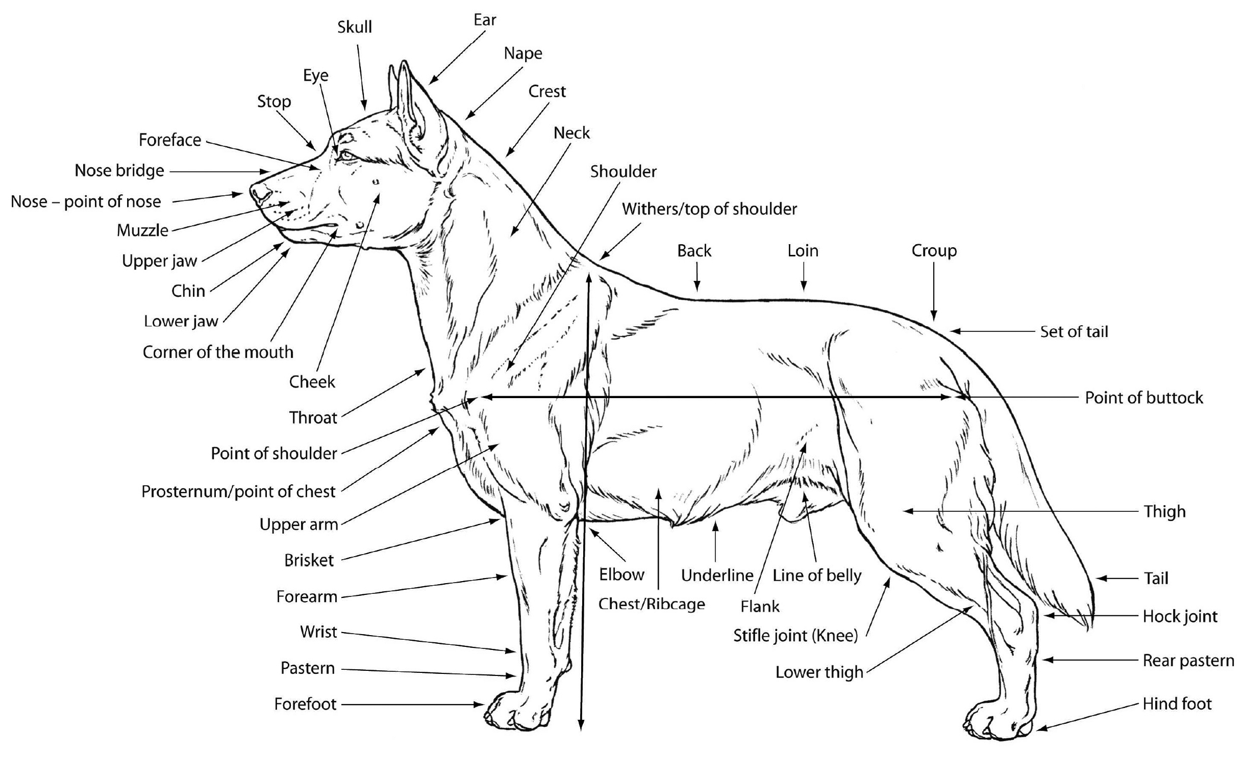
dogexternalanatomy ESL Buzz
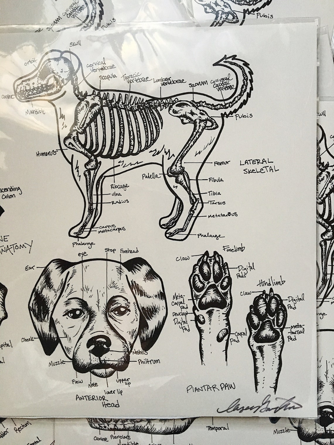
Dog skeleton. As with any vertebrate animal, the skeleton of a dog has the function of supporting the body for movement and protecting its internal organs. We can divide the canine skeleton into three main sections: Axial skeleton: skull, spine, ribs and sternum bones. Appendicular skeleton: bones of the extremities.

dog anatomy Dog Care Training Grooming
Miller's Anatomy of the Dog, 4th Edition; Anatomie comparée des mammifères domestiques: Ostéologie - Arthrologie/Myologie - Robert Barone; Illustrated Veterinary Anatomical Nomenclature. Oskar Schaller, Gheorghe M. Constantinescu. Georg Thieme Verlag, 2007; Nomina Anatomica Veterinaria -6th Edition - 2017; Veterinary Anatomy of Domestic.

Поверхностные мышцы собаки Dog Muscles Anatomy Anatomía del perro, Anatomía animal, Anatomia
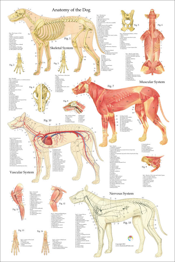
Anatomy atlas of the canine general anatomy: fully labeled illustrations and diagrams of the dog (skeleton, bones, muscles, joints, viscera, respiratory system, cardiovascular system). Positional and directional terms, general terminology and anatomical orientation are also illustrated.

Глубокие мышцы, внутренние органы собаки Dog Muscles & Internal Anatomy Собаки, Животные
Anatomic Planes. The main planes of motion for dogs are as follows (see Figure 5-1): • The sagittal plane divides the dog into right and left portions. If this plane were in the midline of the body, this is the median plane or median sagittal plane. • The dorsal plane divides the dog into ventral and dorsal portions.

Canine Anatomy, Complete Set of 3 Charts. Buy The Set and Save! Industrial
Dog anatomy comprises the anatomical studies of the visible parts of the body of a domestic dog.Details of structures vary tremendously from breed to breed, more than in any other animal species, wild or domesticated, as dogs are highly variable in height and weight. The smallest known adult dog was a Yorkshire Terrier that stood only 6.3 cm (2.5 in) at the shoulder, 9.5 cm (3.7 in) in length.
Canine Dog Anatomy Diagram Print Poster Vet Tech Etsy Ireland
This veterinary anatomy module contains 608 illustrations on the canine myology. Here are presented scientific illustrations of the canine muscles and skeleton from different anatomical standard views (lateral, medial, cranial, caudal, dorsal, ventral / palmar.). Some fascias, tendons, ligaments, joints were labeled.
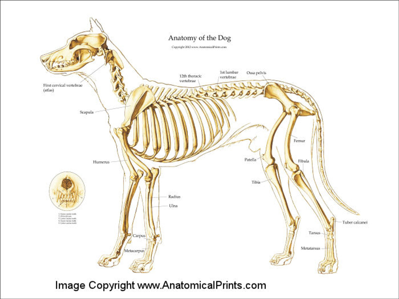
Canine Skeleton Poster Clinical Charts and Supplies
A dog's skeleton is made up of many different bones, which provide structure and support for their body. Dogs have over 300 bones in their body, which is more than humans who have around 206 bones. Their skeleton includes their skull, spine, ribcage and limbs. Dogs have four legs that are designed to help them move quickly and efficiently.

PetMassage Chart 3 Skeleton of the Dog · PetMassage™ Training and Research Institute
It provides information about a dog's skeletal, reproductive, internal, and external anatomy, along with accompanying labeled diagrams. After mating, dogs experience something called a copulatory tie, wherein they remain in the coital position. The male dog dismounts the female at this time. The dogs can remain in this position from a few.
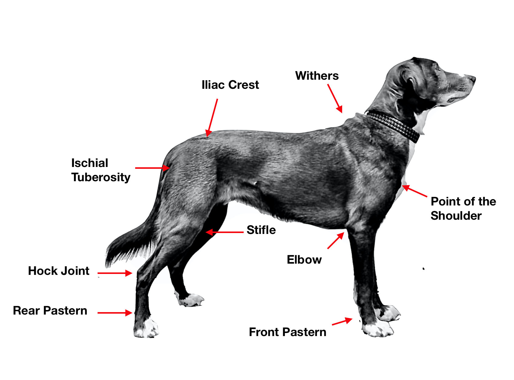
Canine Anatomy 101 Canine Fitness Innovations
Dog Leg Anatomy with Labeled Diagram - Bones, Joints, Muscles and Vessels. 25/04/2023 09/07/2021 by Sonnet Poddar. Most first-year veterinary students have a misconception of the term "leg." Anatomically, the term leg means the part of the hind limb that extends from the stiffle joint to the hock joint (knee to ankle or tibia and fibula.
Dog Anatomy Poster 24 x 36
Dog leg anatomy with the labeled diagrams, Dog sciatic or ischiatic nerve anatomy with a labeled diagram, The fibular nerve lies deep in the thin terminal part of the biceps femoris muscle. This nerve runs directly distal, obliquely crossing the lateral head of the gastrocnemius muscle. It sends an auricular branch to the lateral collateral.

Anatomy and Physiology Canine Muscles Diagram Quizlet
Dog tongue anatomy with special features and labeled diagrams. Dog teeth anatomy. The teeth of a dog are the highly specialized structure in its mouth. You will find forty-two teeth in the mouth cavity of a dog. It is very important to know the eruption time of the dog's permanent teeth a veterinarian.

Canine muscles wccrtv pamppllc caninemarketing petinfographics
The vertebrae of the dog skeleton anatomy also possess some exceptional osteological features that a ruminant or horse. Now, you should try to identify all the bones from the dog skeleton from the actual samples of your anatomy laboratory. You may use the dog bones labeled diagram from the anatomy learner.
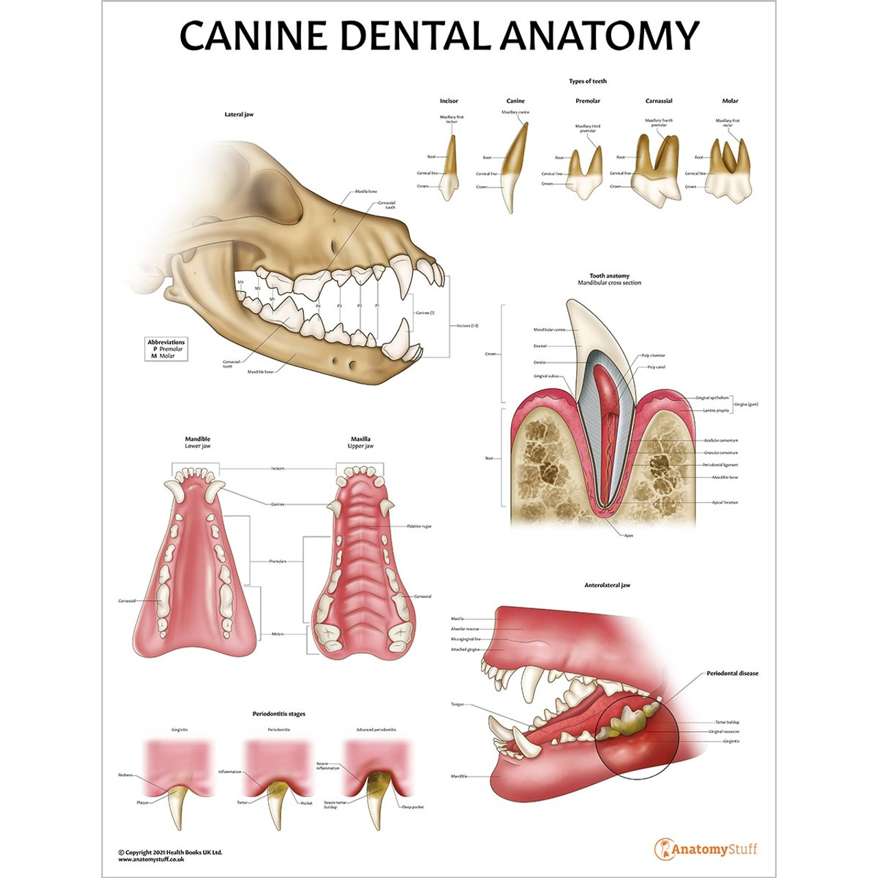
Canine Dental Anatomy Chart Dog Teeth Jaw Poster
The Anatomage Table Vet brings realism into veterinary education by providing an absolutely explicit 3D visualization of animal anatomy. The platform features 3D animal bodies - which include the world's most detailed canine cadaver - that assist anatomy inspection. Introduce a high-quality approach to animal learning using digital.
M. Douglas Wray Dog Anatomy
Heart. A dogs heart beats between 70 and 120 times per minute, compared to a humans 70 - 80 beats per minute. Dogs take between 10 and 30 breathes every minute. Dogs have a visual range of 250 degrees compared to the human range of 180 degrees. A dogs temperature is between 100.2 and 102.8 degrees Fahrenheit.

2021 Ultimate Guide to Dog Anatomy. As the pace of veterinary advancement accelerates, even the most experienced veterinary teams are challenged to keep up with all the changes that impact their practice.. potential outcomes and prognoses and tools e.g. anatomical diagrams, clinical formulas and programs and home care videos; Enhancing the.
