
Cross Section Of The Human Eye
Media in category "Human eyeball cross-section" The following 131 files are in this category, out of 131 total. 007alhazen eye.jpg 342 × 512; 38 KB. 1413 Structure of the Eye.jpg 2,175 × 1,242; 1.06 MB. 1490-95 da vinci - codex atlanticus.jpg 580 × 870; 157 KB. 201405 eye.png 400 × 400; 47 KB.
Human eye anatomy with cross section of eye diagram 1783141 Vector Art at Vecteezy
Eye in Cross Section. Click on a label to display the definition. Tap on the image or pinch out and pinch in to resize the image.

Human Eye Anatomy 3D Model Cross Section with all Eye parts Ready for Renderings, 3D Printings
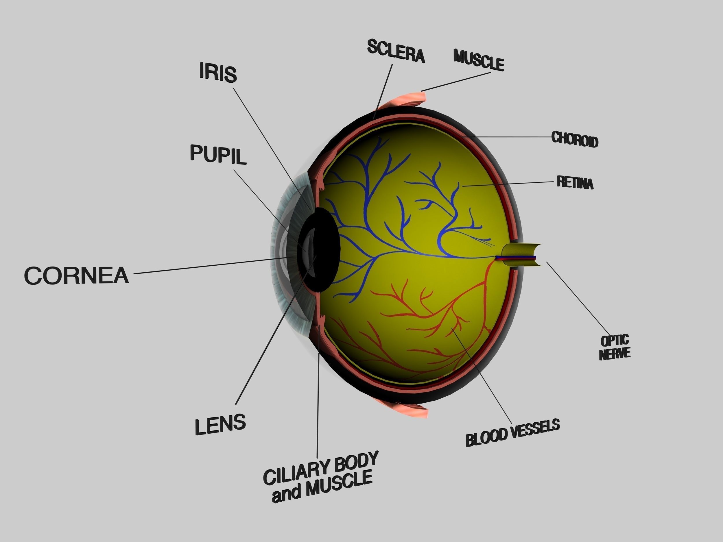
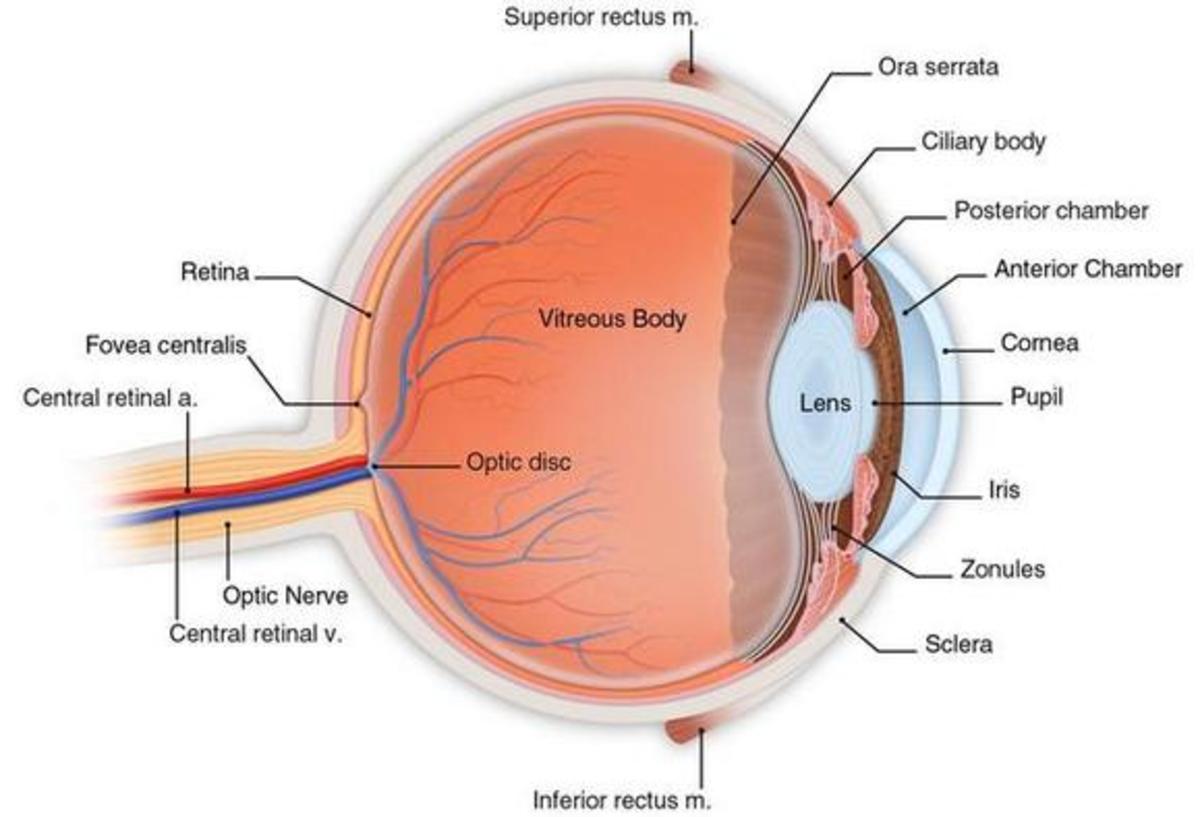
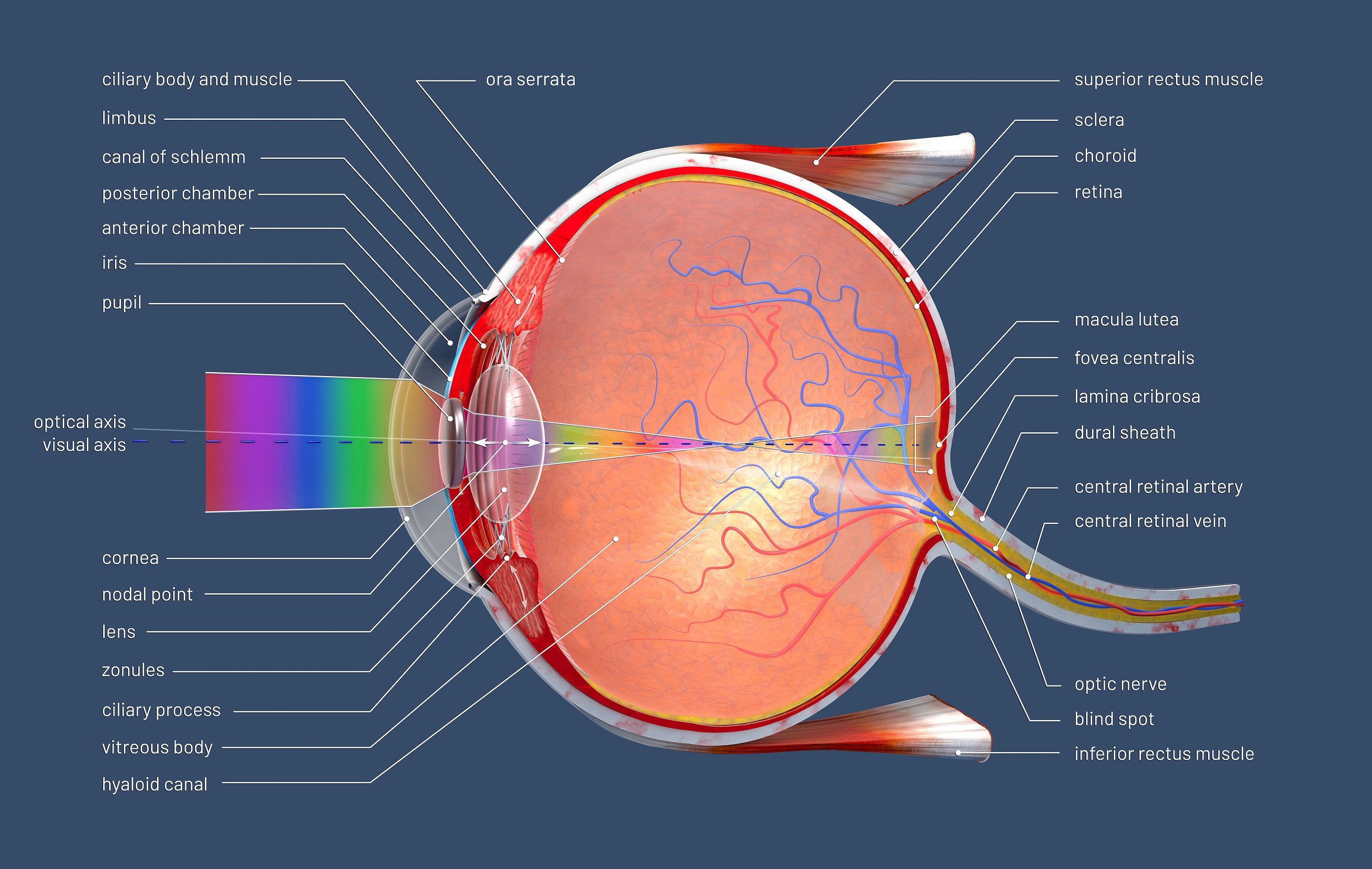
Behind the anterior chamber is the eye's iris (the colored part of the eye) and the dark hole in the middle called the pupil. Muscles in the iris dilate (widen) or constrict (narrow) the pupil to control the amount of light reaching the back of the eye. Directly behind the pupil sits the lens. The lens focuses light toward the back of the eye.

Cross Section Of The Human Eye
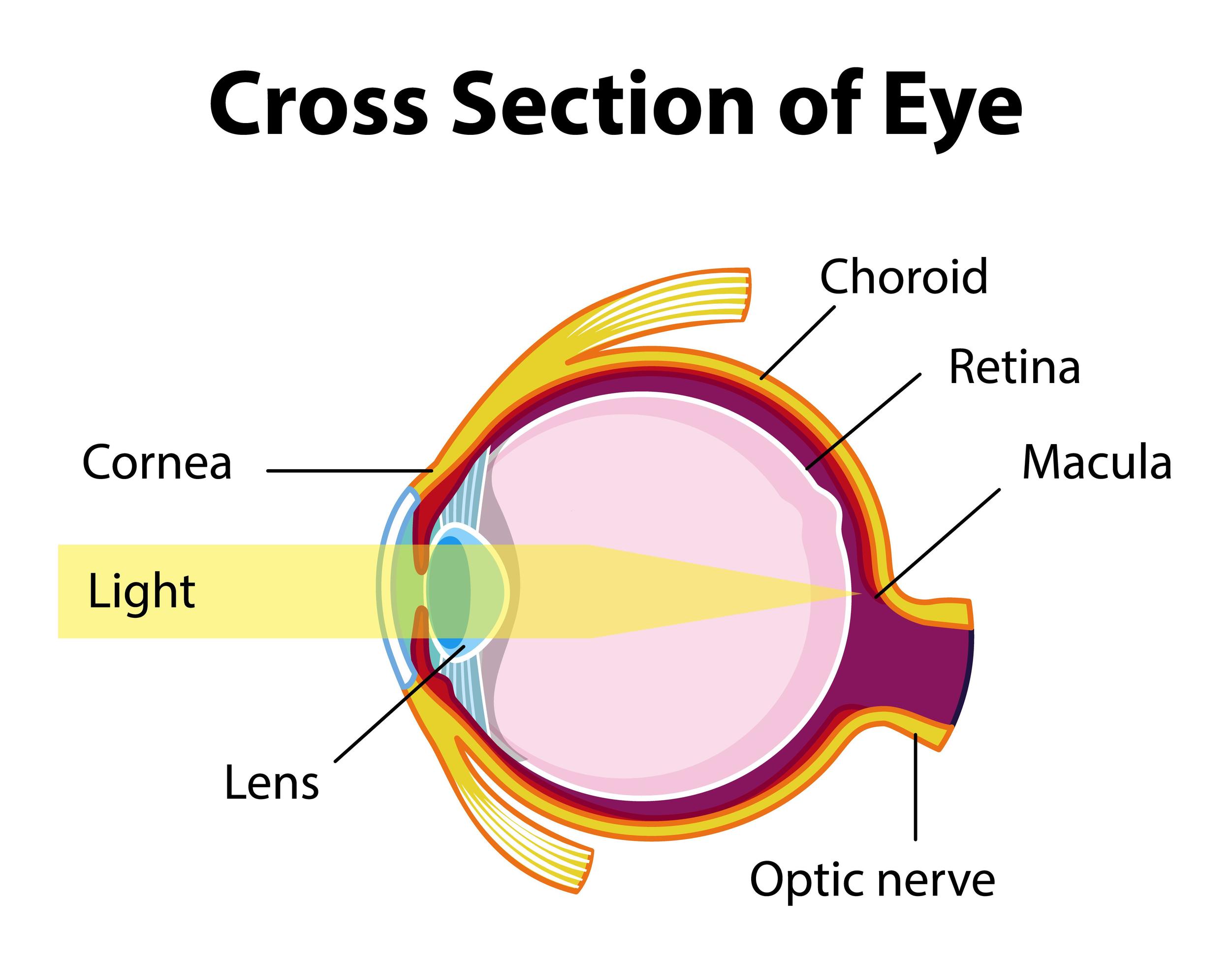
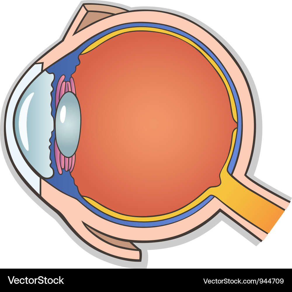
Cross Section of an EyeballThe eyeball has three major coats-the smooth, protective outer sclera, the middle, pigmented choroid and the inner, light-sensitiv.
Human Eye Cross Section Eyeball 3D Model OBJ 3DS FBX C4D DXF STL
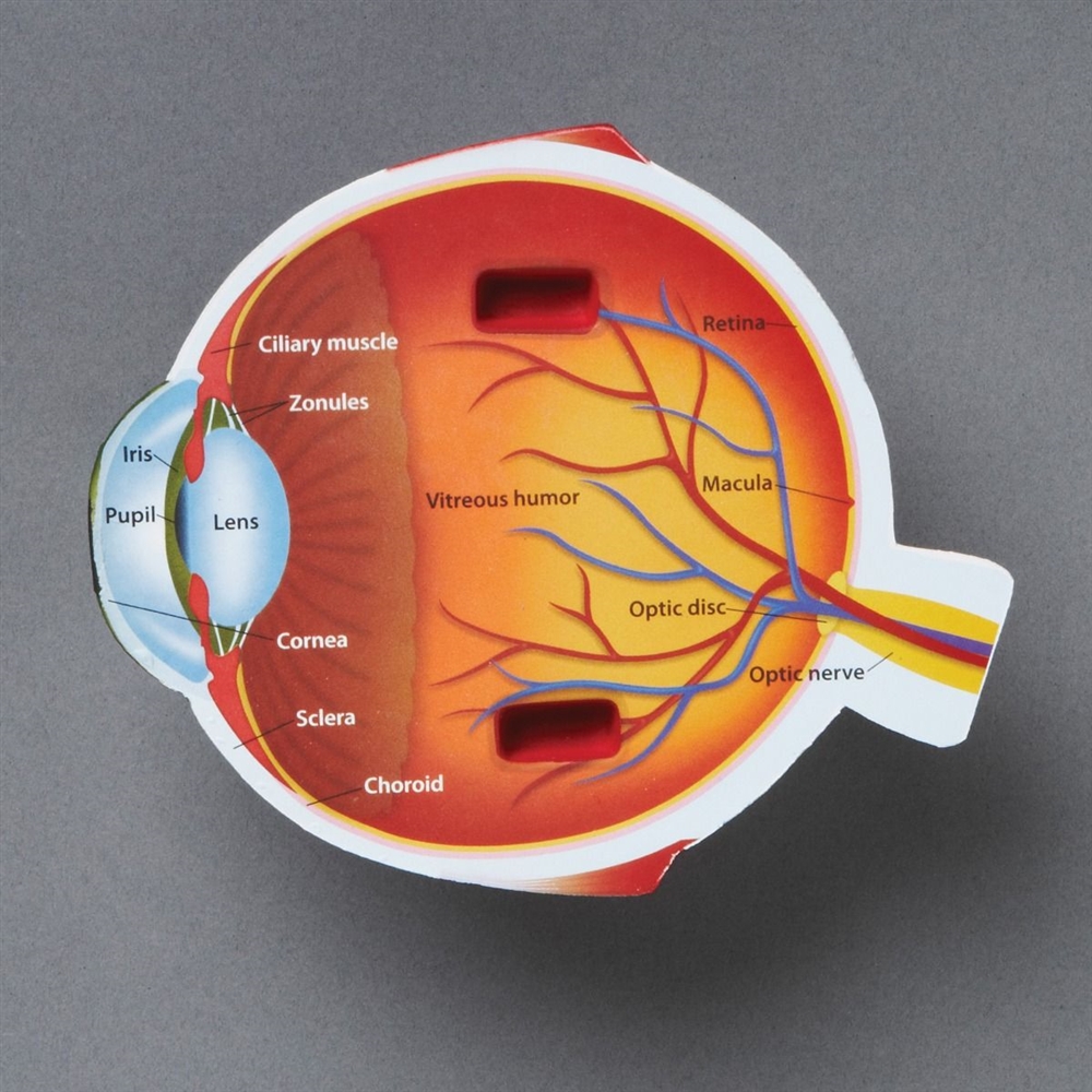
Cross-section of the eye. The zonules of Zinn keep the lens suspended, and the muscles of the ciliary body focus the lens. The ciliary body also secretes aqueous humor, which fills the anterior and posterior chambers, passes through the pupil into the anterior chamber, and drains primarily via the Schlemm canal. The iris regulates the amount of.
Montessori Materials Cross Section Eye Model
Cross-section of the eye The zonules of Zinn keep the lens suspended, and the muscles of the ciliary body focus the lens. The ciliary body also secretes aqueous humor, which fills the anterior and posterior chambers, passes through the pupil into the anterior chamber, and drains primarily via Schlemm's canal.

Eye Cross Sectional Anatomy Michelle Davis Studios
Representation of a horizontal section of the eyeball reveals its three coats: (1) external or fibrous coat (sclera and cornea); (2) middle or vascular coat (choroid, ciliary body, and iris); and (3) internal or retinal layer. The four refractive media are the cornea, the aqueous humor in the anterior chamber, the lens, and the vitreous body.
Human eye ball crosssection 3D model TurboSquid 1376280
On the cross-section of the eye, we can identify the two chambers of the eyeball filled with the aqueous humor; anterior and posterior. The anterior chamber of eyeball is found between the cornea and iris. The posterior chamber of eyeball is more of a slit-like cavity, found between the iris and lens. Fascial sheath (Tenon's capsule)
Anatomy of the Eye Human Eye Anatomy Owlcation
Cross-section of the eye. The zonules of Zinn keep the lens suspended, and the muscles of the ciliary body focus the lens. The ciliary body also secretes aqueous humor, which fills the anterior and posterior chambers, passes through the pupil into the anterior chamber, and drains primarily via the Schlemm canal. The iris regulates the amount of.

Cross Section Of Human Eye Digital Art by Stocktrek Images Pixels
Human eye anatomy diagram, medical illustration. Isolated on a white bacground. Eye anatomy. Rod cells and cone cells. Eye anatomy. Rod cells and cone cells. The arrangement of retinal cells is shown in a cross section. Vector diagram for your design, educational, biological, science and medical use.

Human Eye Cross Section Eyeball 3D Model OBJ 3DS FBX C4D DXF STL
This is a cross-sectional study of patients with clinically-significant dry eye, suspected glaucoma (controls), glaucoma, or cataracts.. The ROC curve constructed from the final model's ability to predict a diagnosis of dry eye from glaucoma had a cross-validated mean AUC of 0.93 (cross-validated SD = 0.026), with a sensitivity of 89% and.

Crosssection through the human eye — Science Learning Hub
human eye, in humans, specialized sense organ capable of receiving visual images, which are then carried to the brain.. Anatomy of the visual apparatus Structures auxiliary to the eye The orbit. The eye is protected from mechanical injury by being enclosed in a socket, or orbit, which is made up of portions of several of the bones of the skull to form a four-sided pyramid, the apex of which.

Human Eye Cross Section Eyeball 3D Model OBJ 3DS FBX C4D DXF STL
Cross-section of the eye. The zonules of Zinn keep the lens suspended, and the muscles of the ciliary body focus the lens. The ciliary body also secretes aqueous humor, which fills the anterior and posterior chambers, passes through the pupil into the anterior chamber, and drains primarily via Schlemm's canal. The iris regulates the amount of.

eye cross section Discovery Eye Foundation
Dry eye syndrome is the most common eye disease, and if untreated, it can cause corneal ulcerations, scarring, and even perforation.. is an approximately 1.5mm area of specialized avascular retina that can be identified as a depression in the retina in cross-section. The foveola is the central floor of the fovea, approximately 0.35mm in.
Human eye cross section Royalty Free Vector Image
A cross-sectional view of the eye shows : Three different layers: The external layer, formed by the sclera and cornea. The. The sagittal section of the eye also reveals the lens, which is a transparent body located behind the iris. The lens is suspended by ligaments (called zonule fibers) attached to the anterior portion of the ciliary body..
3d illustration of a cross section of the human eye with explanations and inscription Stephan
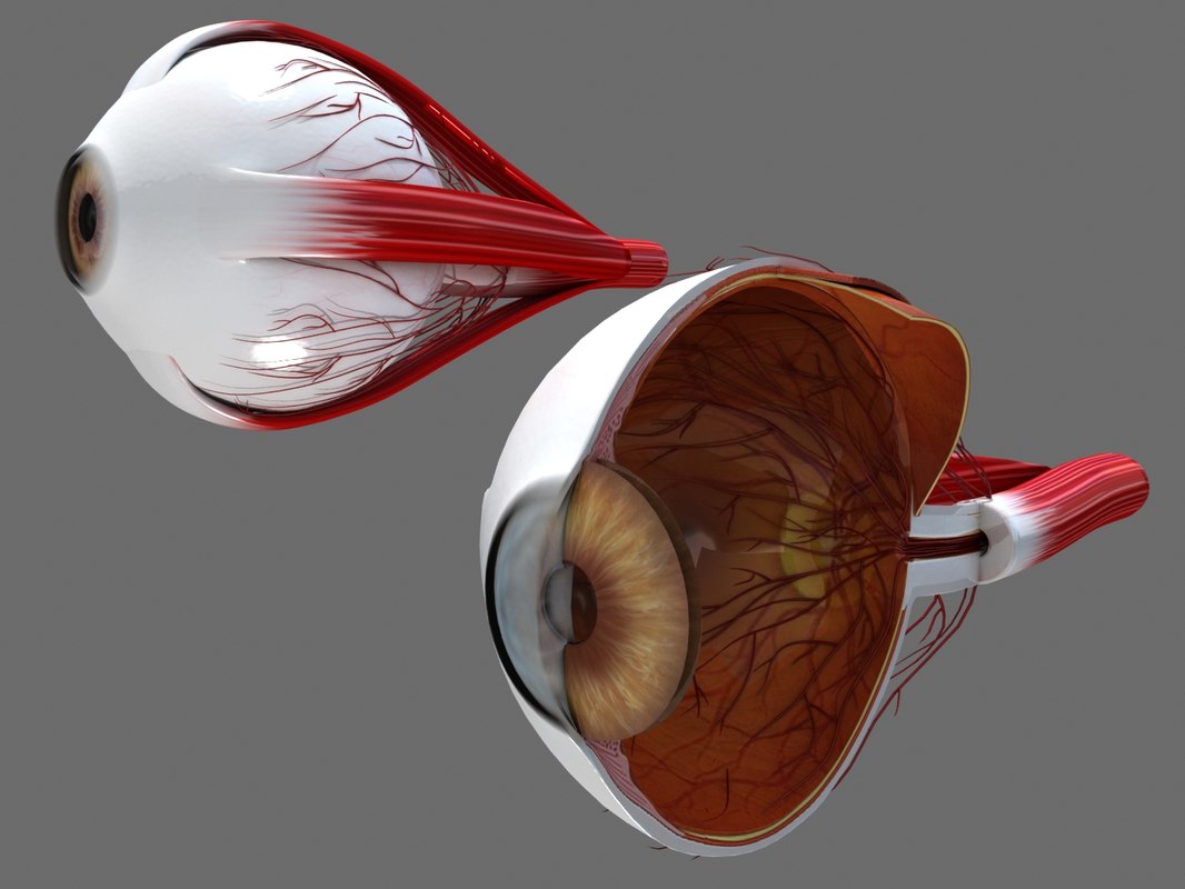
Cross section of the human eyeball viewed from above. Dave Carlson / CarlsonStockArt.com. Iris. The choroid continues at the front of the eyeball to form the iris. The iris is a flat, thin, ring-shaped structure sticking into the anterior chamber. This is the part that identifies a person's eye colour.
