Dog Internal Anatomy Anatomical Charts & Posters
Dog anatomy comprises the anatomical studies of the visible parts of the body of a domestic dog.Details of structures vary tremendously from breed to breed, more than in any other animal species, wild or domesticated, as dogs are highly variable in height and weight. The smallest known adult dog was a Yorkshire Terrier that stood only 6.3 cm (2.5 in) at the shoulder, 9.5 cm (3.7 in) in length.
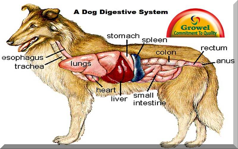
How is a Dog Digestive System Functioning? Growel Agrovet
What is dog anatomy. Dog anatomy is how a dog is built. In other words, a dog's anatomy includes all the parts of a dog's body like: Skeletal structure. Internal organs. Musculoskeletal system. Senses. Body systems. Each body part plays an important role in how your dog moves, breathes, eats, and reproduces.

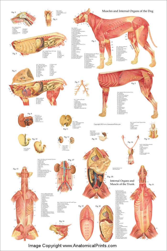
A4 Veterinary Poster u00 Internal Organs Of The Dog (Animal Anatomy Pathology) Dog anatomy
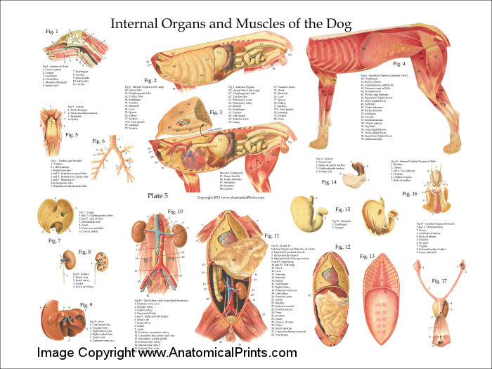
This detailed canine internal anatomy wall chart has been laminated for easy cleaning and to enable wipeable marker pens to be used for notation. This is one of our bestselling veterinary charts in the canine anatomy series, which includes the canine muscular system and canine skeletal anatomy charts. Designed and printed in the UK.
Dog Digestive Process and what the stages are and how it works
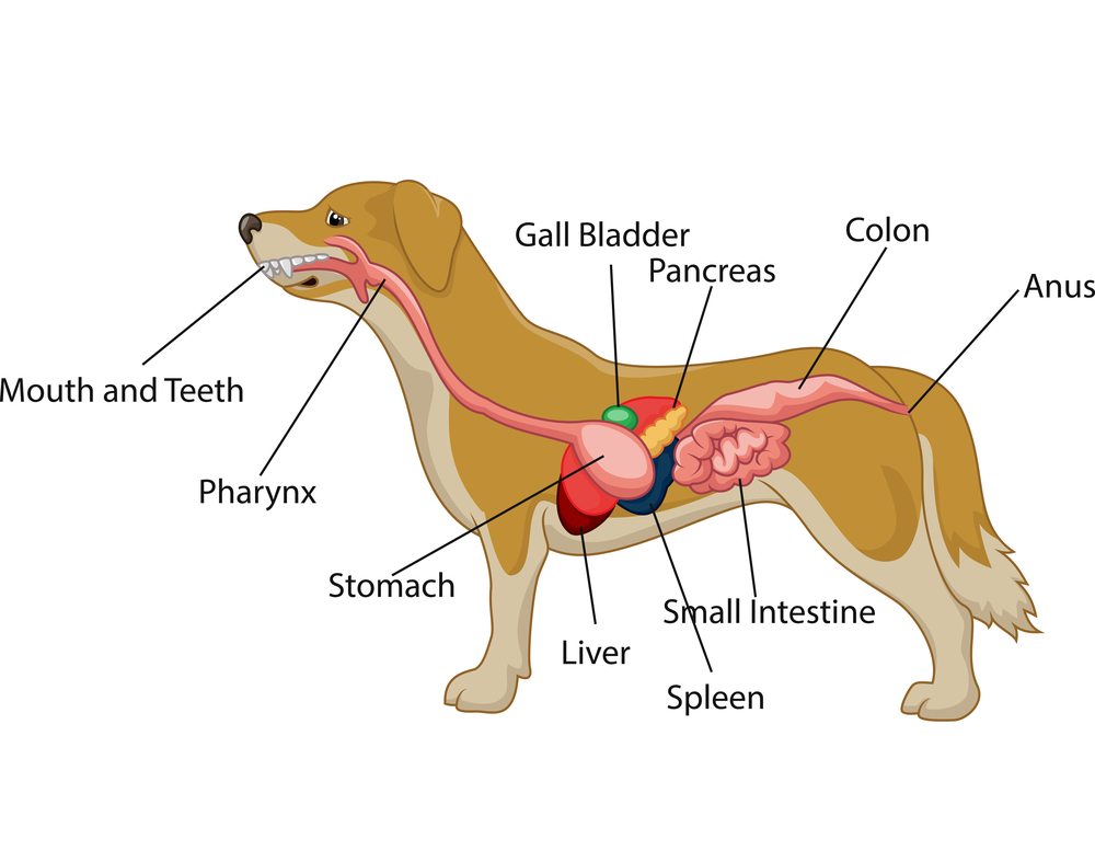
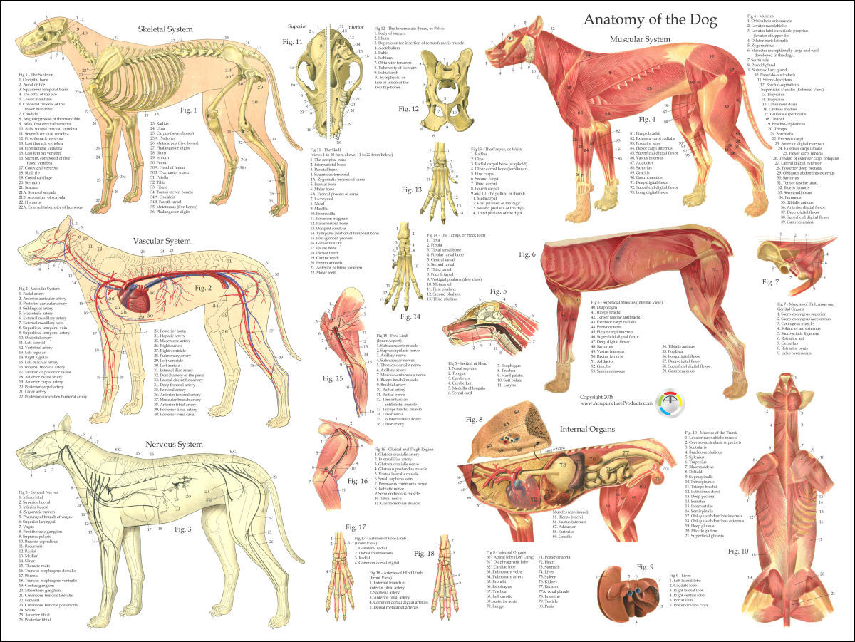
Quick idea: in this article, you will learn the location of different organs from the different systems (like skeletal, digestive, respiratory, urinary, cardiovascular, endocrine, nervous, and special sense) of a dog with their important anatomical features.
Dog Internal Anatomy Poster 24 x 36
A dog's physical anatomy is designed to help them navigate their environment and perform various tasks. Their bodies are made up of many different parts, including their skeleton, muscles and internal organs. One of the most important parts of a dog's anatomy is their skeleton.

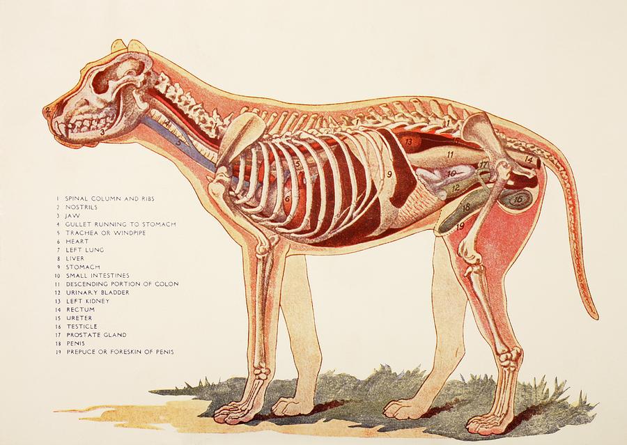
Anatomy of a male dog crosssection, showing the skeleton and internal organs. Colour process
Xiphoid region (Cranial abdominal region) Zygomatic bone. Zygomatic gland. Zygomatic region. Radiographic anatomy: labeled images in the transverse plane of a healthy dog's whole body, using tomodensitometry. Introduction to the anatomy of the skull, thorax, abdomen, pelvic cavity, muscles and blood vessels: main anatomical structures identified.
Dog Anatomy (Thoracic and Abdominal Organs)
Internal Anatomy of the Female Dog's Body Female vs. Male Dog Anatomy Comparison Health Considerations Conclusion FAQs External Anatomy of Female Dogs The female dog anatomy bears features both common and unique to her gender. Observing them helps in general care and detecting health abnormalities.

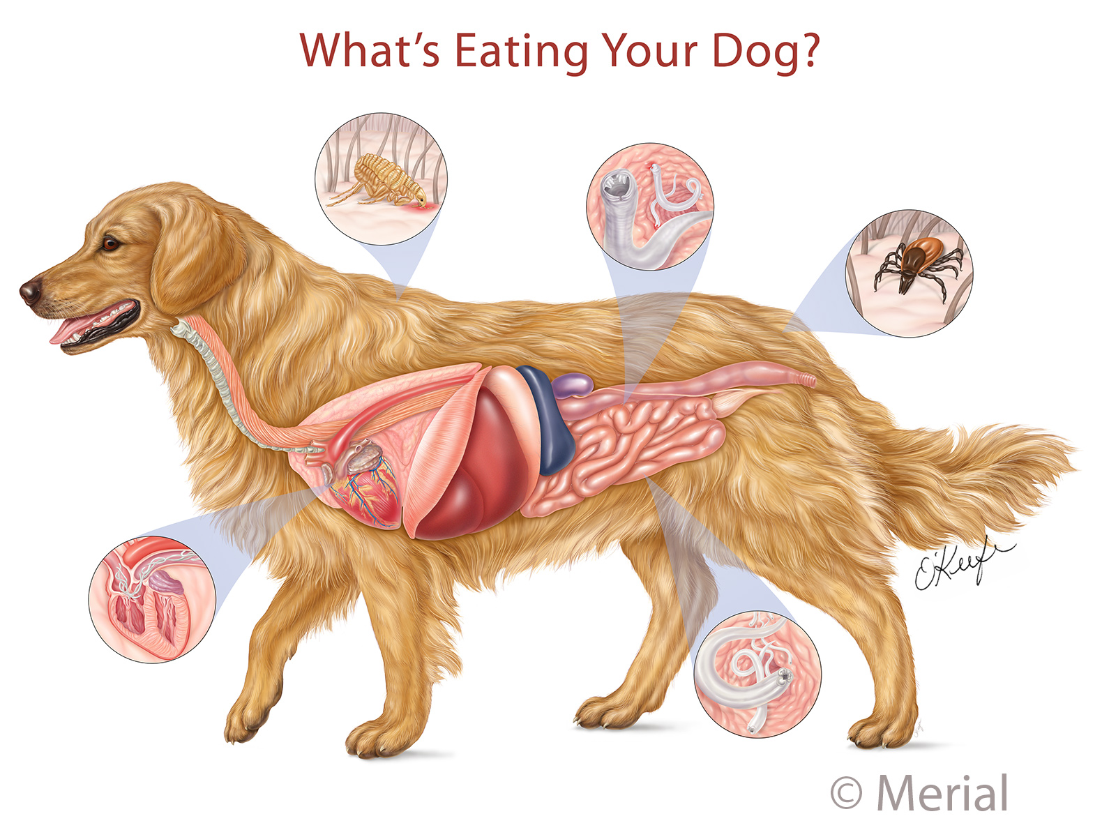
Dog anatomy4 views Illustration by Laurie O'Keefe Medical Illustration & Animation
Our canine charts cover internal organ anatomy, the musculoskeletal system, common pathologies and guides to dog health and safety. Excellent wall displays in vet clinics, surgeries, dog groomers, and veterinary colleges. Our canine posters are suitable for both animal lovers and veterinary studies. Our canine model range covers detailed.

Digestive system of the dog Royalty Free Vector Image
Common anatomical terminology Here are some common veterinary terms and their meanings: Pet senses Pets communicate in a very different way than people do. They have the same basic senses like sight, hearing, smell, touch, and taste, but they use them differently to communicate with the world.

Глубокие мышцы, внутренние органы собаки Dog Muscles & Internal Anatomy Собаки, Животные
Speaking of skeletons, a dog has 320 bones in their body (depending on the length of their tail) and around 700 muscles. Muscles attach to bones via tendons. Depending on the breed of dog, they will have different types of muscle fibers. You've probably heard about slow and fast twitch muscle fibers before.
Dog Internal Anatomy Poster
In addition to the world's most segmented dog anatomy, the Table Vet also includes a diverse library of animal cases.. The Anatomage Dog is the first highly detailed dog anatomy atlas that comprehensively features internal organs, including vascular systems and muscular-skeletal structures. Originating from real dog data, the Anatomage Dog.
Dog Muscle Skeletal Veterinary Internal Anatomy Poster 18 X 24 Laminated Chart Art Posters
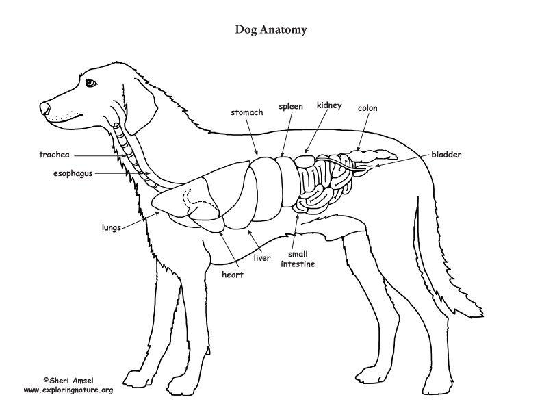
Internal anatomy of a dog: carnivorous domestic mammal raised to perform various tasks for humans. Encephalon: seat of the intelluctual capacities of a gog. Spinal column: important part of the nervous system. Stomach: part of the digestive tract between the esophagus and the intestine. Spleen: hematopoiesis organ that produces lymphocytes.

Dog Anatomy Skeleton Animaltia
We discuss the internal and external anatomy of dogs so that you can see that, despite individual differences, there is a reason they are all considered part of the same species. You may also be interested in: Anatomy of a Frog - Internal and External Contents Canine anatomy Dog skeleton Muscles of the dog Organs of dogs Canine anatomy

Dog anatomy Royalty Free Vector Image VectorStock
Dogs, like all mammals, have eyes, a nose, a forehead, and ears. The only difference is that their noses are cold and wet, and their ears can be either dropped, erect, or cropped, depending on the breed. They also have a throat, a flew (the upper lip), chest, fore and hind legs, back, stomach, buttocks, and a tail.
Internal Organs Of A Male Dog. From Photograph by Ken Welsh Pixels
On the left side view of a dog's internal organs, you can see the lungs, heart, liver, stomach, spleen, kidney, intestines, bladder, and the rectum in that order from front to back. You can also view the spinal column and the brain. Laurie O'Keefe Dog Anatomy Organs Right Side
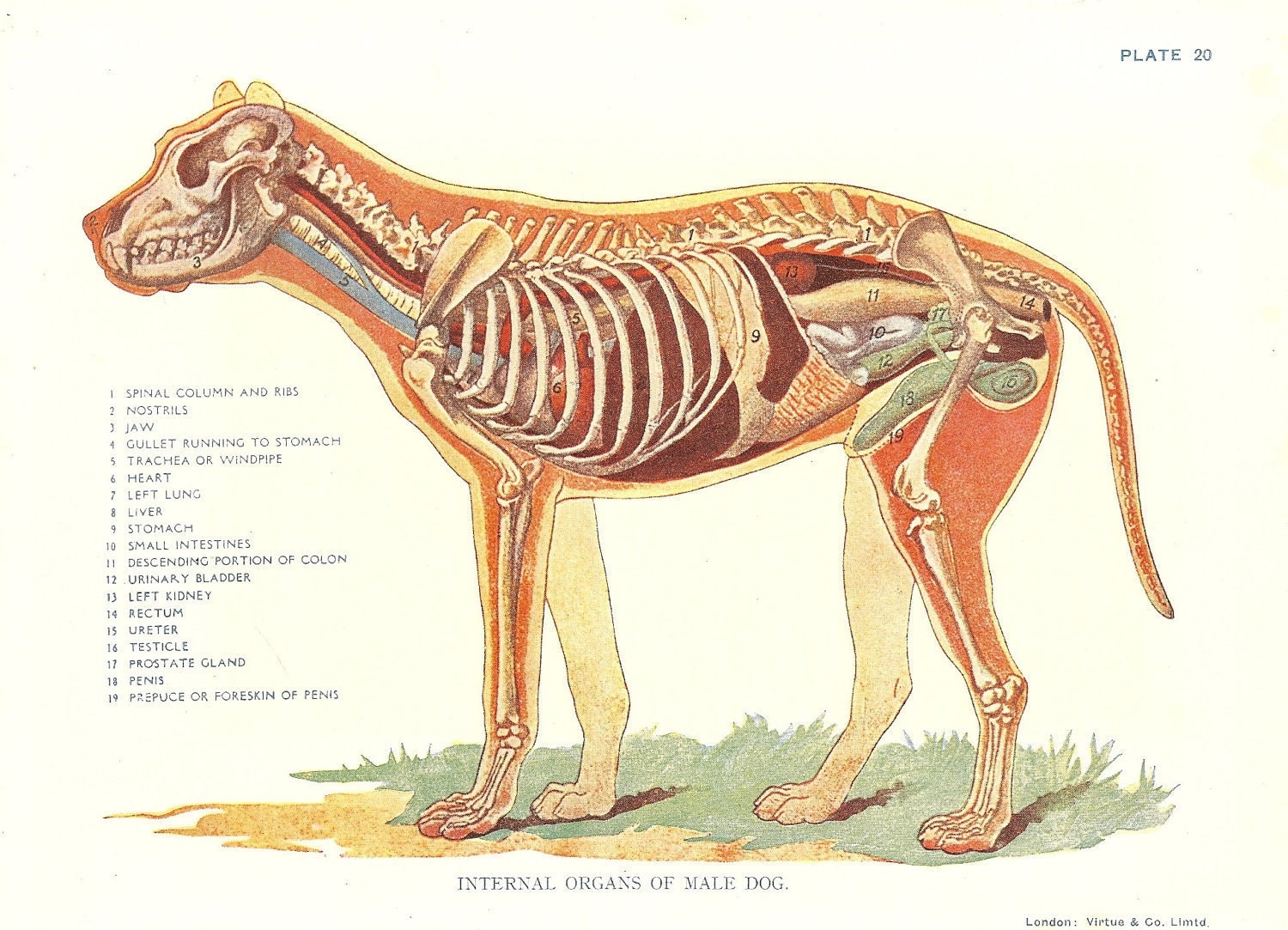
Dog Veterinary Print 1920s Internal Organs Of Male Dog
This module of vet-Anatomy is a basic atlas of normal imaging anatomy of the dog on radiographs. 51 sampled x-ray images of healthy dogs performed by Susanne AEB Borofka (PhD - dipl. ECVDI, Utrecht, Netherland) were categorized topographically into seven chapters (head, vertebral column, thoracic limb, pelvic limb, larynx/pharynx, thorax and abdomen/pelvis).
