Pictures Of Bones Of The Feet
Ankle anatomy. The ankle joint, also known as the talocrural joint, allows dorsiflexion and plantar flexion of the foot. It is made up of three joints: upper ankle joint (tibiotarsal), talocalcaneonavicular, and subtalar joints.The last two together are called the lower ankle joint. The upper ankle joint is formed by the inferior surfaces of tibia and fibula, and the superior surface of talus.
Bones of human foot with labels on white background — phalanx, fibula Stock Photo 200635320
2,836 Human Foot Bones Stock Photos, High-Res Pictures, and Images - Getty Images Boards Sign in Browse Creative Images Creative Images Browse millions of royalty-free images and photos, available in a variety of formats and styles, including exclusive visuals you won't find anywhere else. See all creative images Trending Image Searches
Bones of the human foot diagram 1142236 Vector Art at Vecteezy
kool99/Getty Images In the foot, there are: 26 bones 33 joints more than 100 muscles, tendons, and ligaments Bones of the foot The bones in the foot make up nearly 25% of the total.

Foot & Ankle Bones
Biology Foot Bone Human leg Human body Pain Foot Anatomy royalty-free images 59,192 foot anatomy stock photos, 3D objects, vectors, and illustrations are available royalty-free. See foot anatomy stock video clips Filters All images Photos Vectors Illustrations 3D Objects Sort by Popular
3D Human Foot Tarsals and Metatarsal Bones Collection Yellow 12 models TurboSquid 1749962
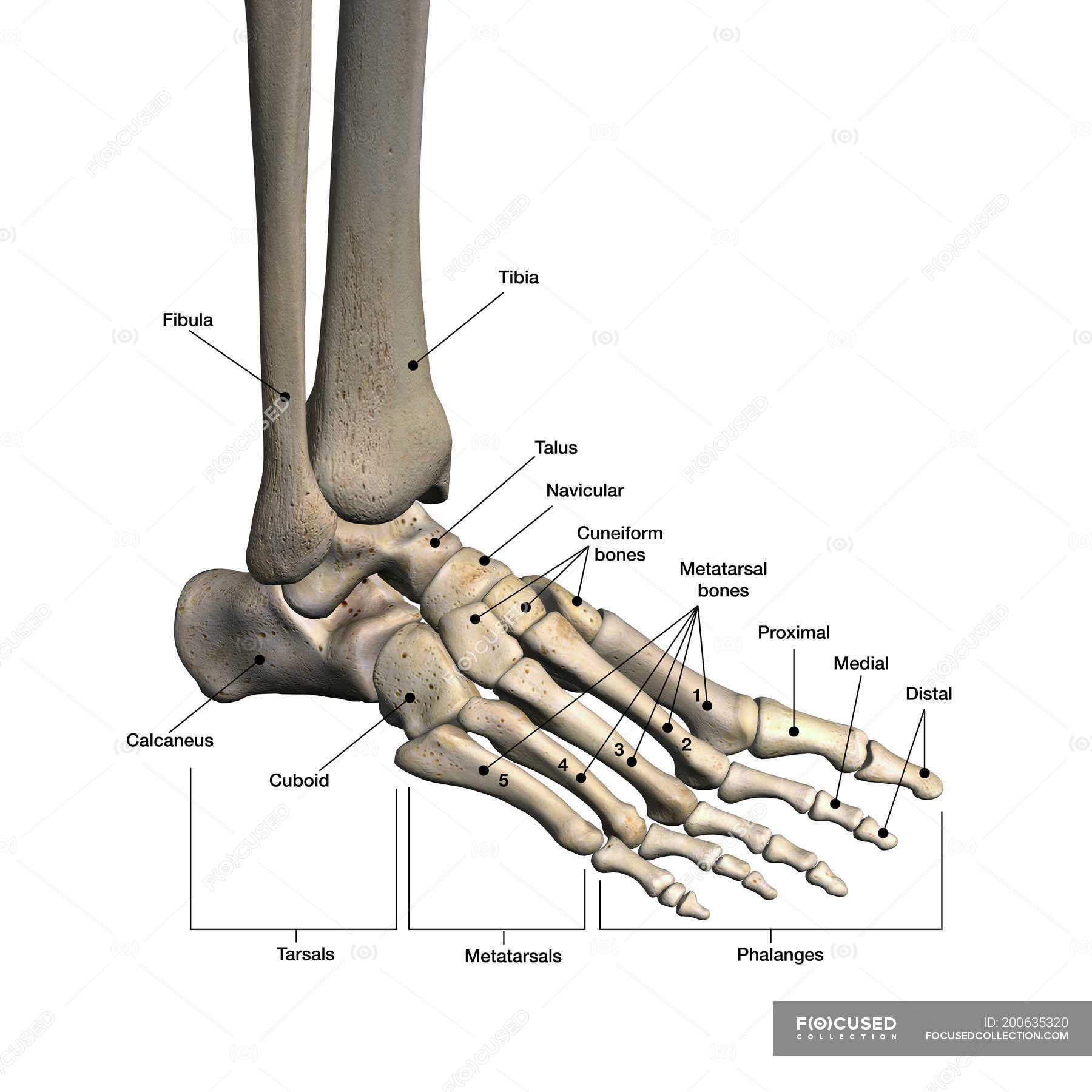
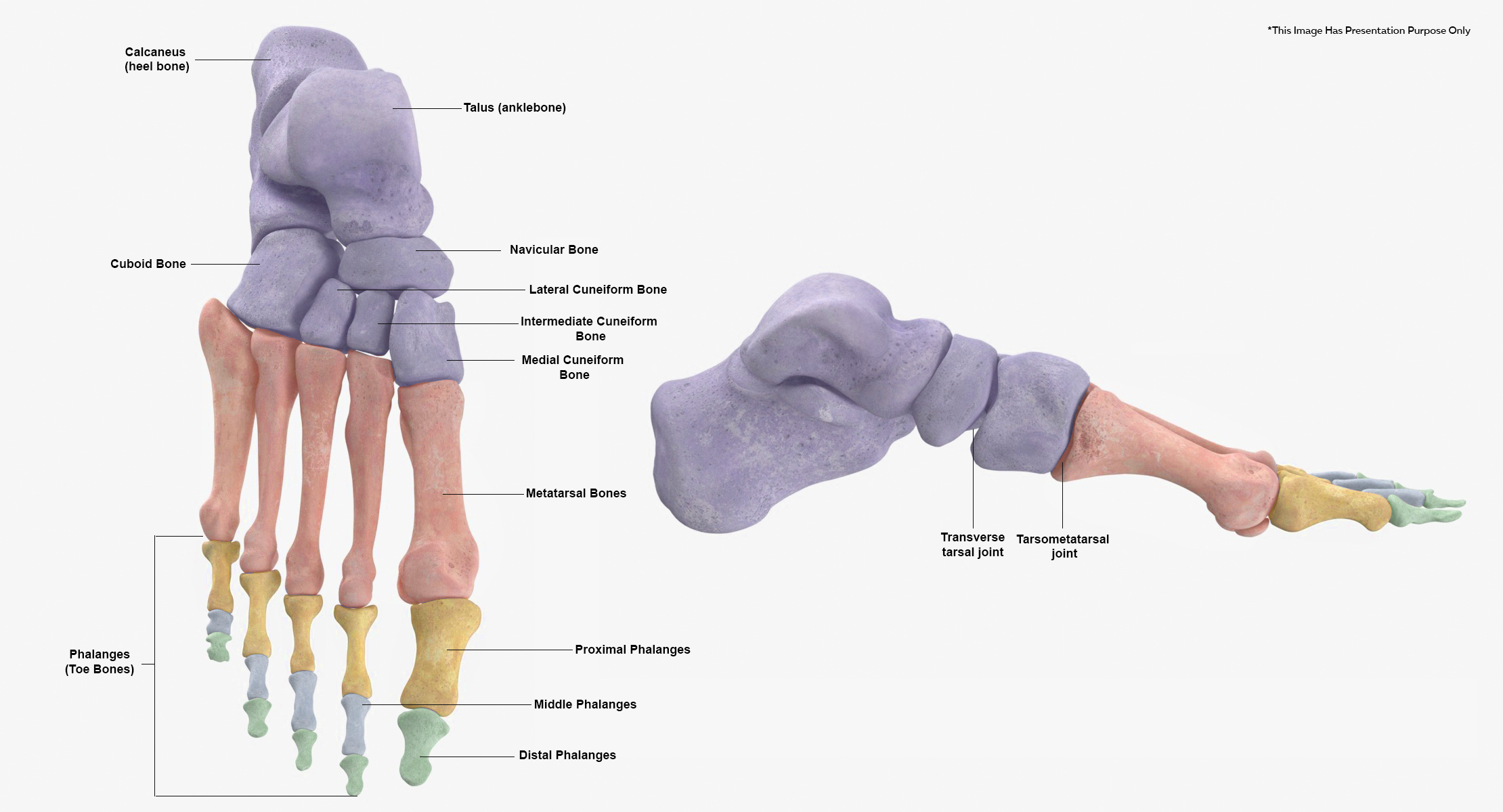
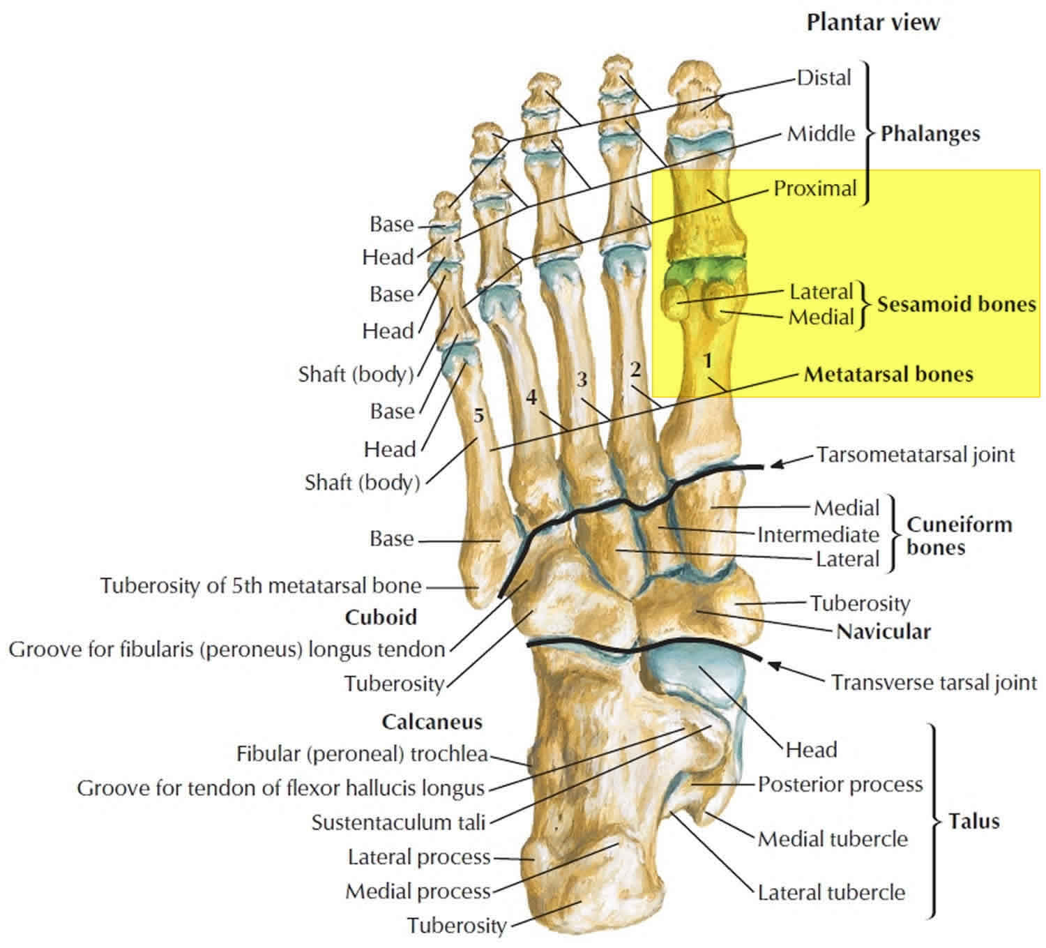
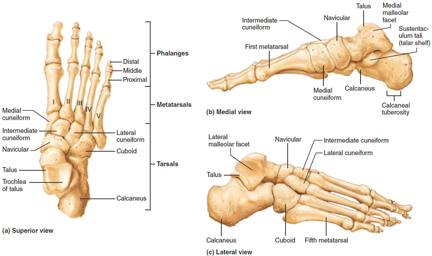
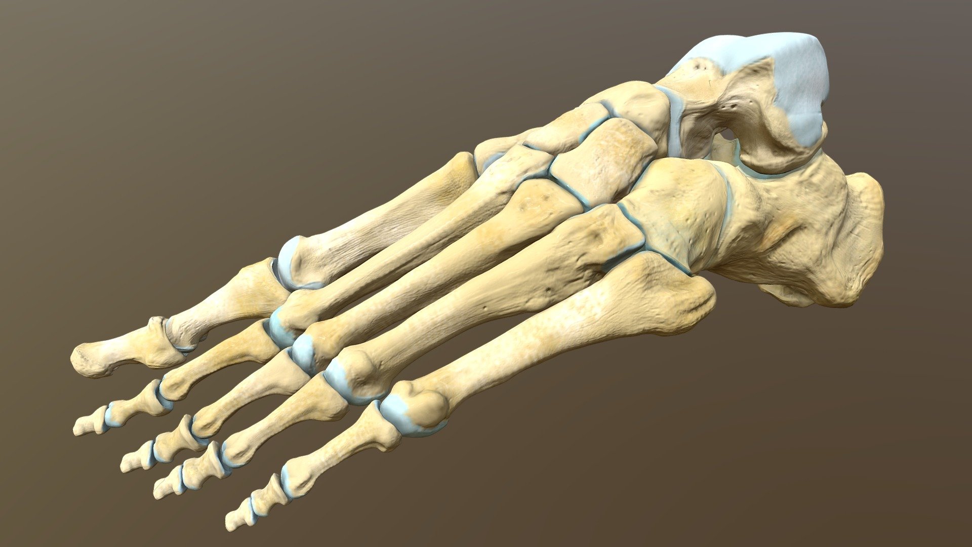
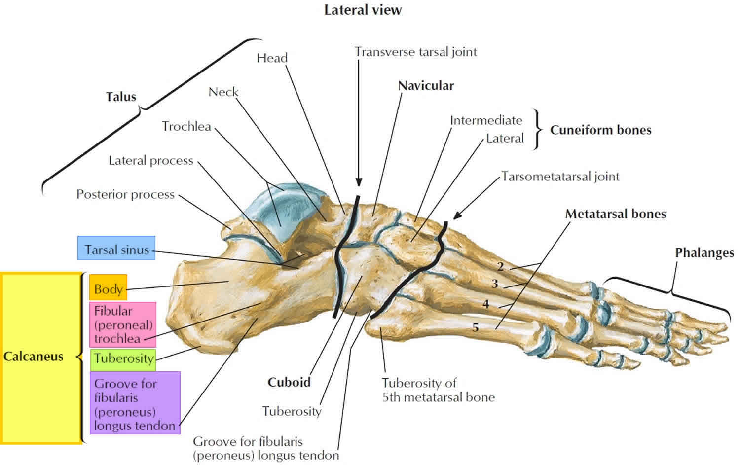
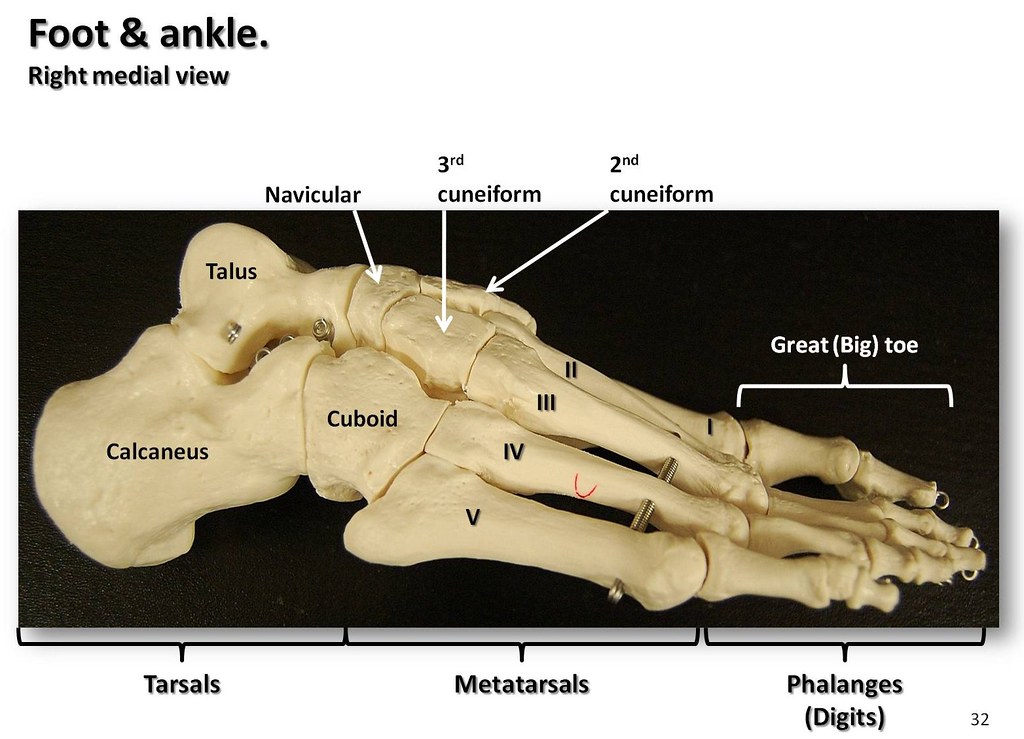
Human body Skeletal System Bones of foot Bones of foot The 26 bones of the foot consist of eight distinct types, including the tarsals, metatarsals, phalanges, cuneiforms, talus,.
Turf toe causes, signs, symptoms, recovery, diagnosis & turf toe treatment
Medial cuneiform Intermediate cuneiform Lateral cuneiform Some people may be born with an extra navicular bone ( accessory navicular) beside the regular navicular bone, on the inside of the foot. This is a normal anatomical variation seen in around 2.5% of the entire population of the US. Metatarsal Bones
Flat Feet Causes In Adults & Children, Symptoms, Exercises & Treatment
Foot Anatomy The foot contains 26 bones, 33 joints, and over 100 tendons, muscles, and ligaments. This may sound like overkill for a flat structure that supports your weight, but you may not realize how much work your foot does!

How to have beautiful, healthy feet banish bunions and other abominations! Harrogate Yoga
58,169 bones of the foot stock photos, vectors, and illustrations are available royalty-free. See bones of the foot stock video clips Image type Orientation People Sort by Geography and Landscapes Healthcare and Medical Anatomy Emotion Biology bone foot ankle skeleton pain
.jpg)
Foot Bone Diagram resource Imageshare
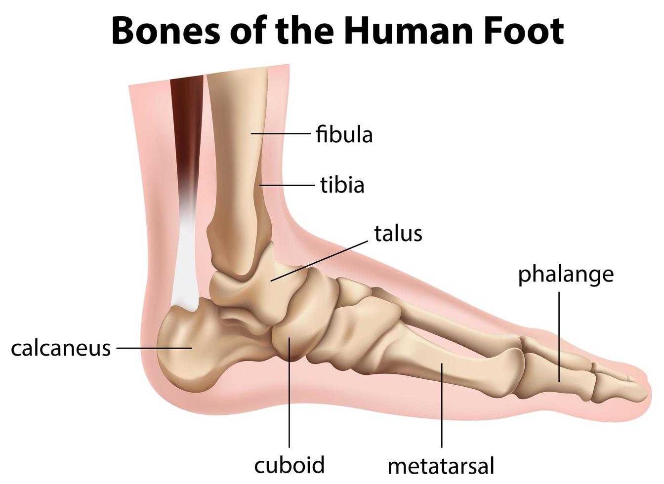
Picture of Foot. The feet are located at the end of the legs and are used to stand and walk. Feet are very complex, comprised of 28 bones and 30 joints. The tendons, ligaments, and muscles in the feet number more than 100. Believe it or not, the feet absorb more than 100,000 pounds of pressure during one mile of walking.
foot bones 4 CoreWalking
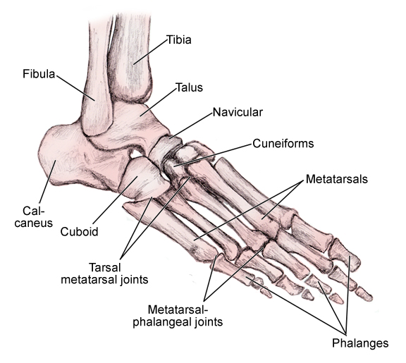
The bones of the foot provide mechanical support for the soft tissues; helping the foot withstand the weight of the body whilst standing and in motion. They can be divided into three groups: Tarsals - a set of seven irregularly shaped bones. They are situated proximally in the foot in the ankle area. Metatarsals - connect the phalanges to.
Left human foot bones Buy Royalty Free 3D model by Catherine Sulzmann (csulzmann) [3665b79
Browse 4,222 foot bones photos and images available, or search for human foot bones to find more great photos and pictures. Browse Getty Images' premium collection of high-quality, authentic Foot Bones stock photos, royalty-free images, and pictures. Foot Bones stock photos are available in a variety of sizes and formats to fit your needs.

Foot Bone Anatomy Vector Illustration 539973 Vector Art at Vecteezy
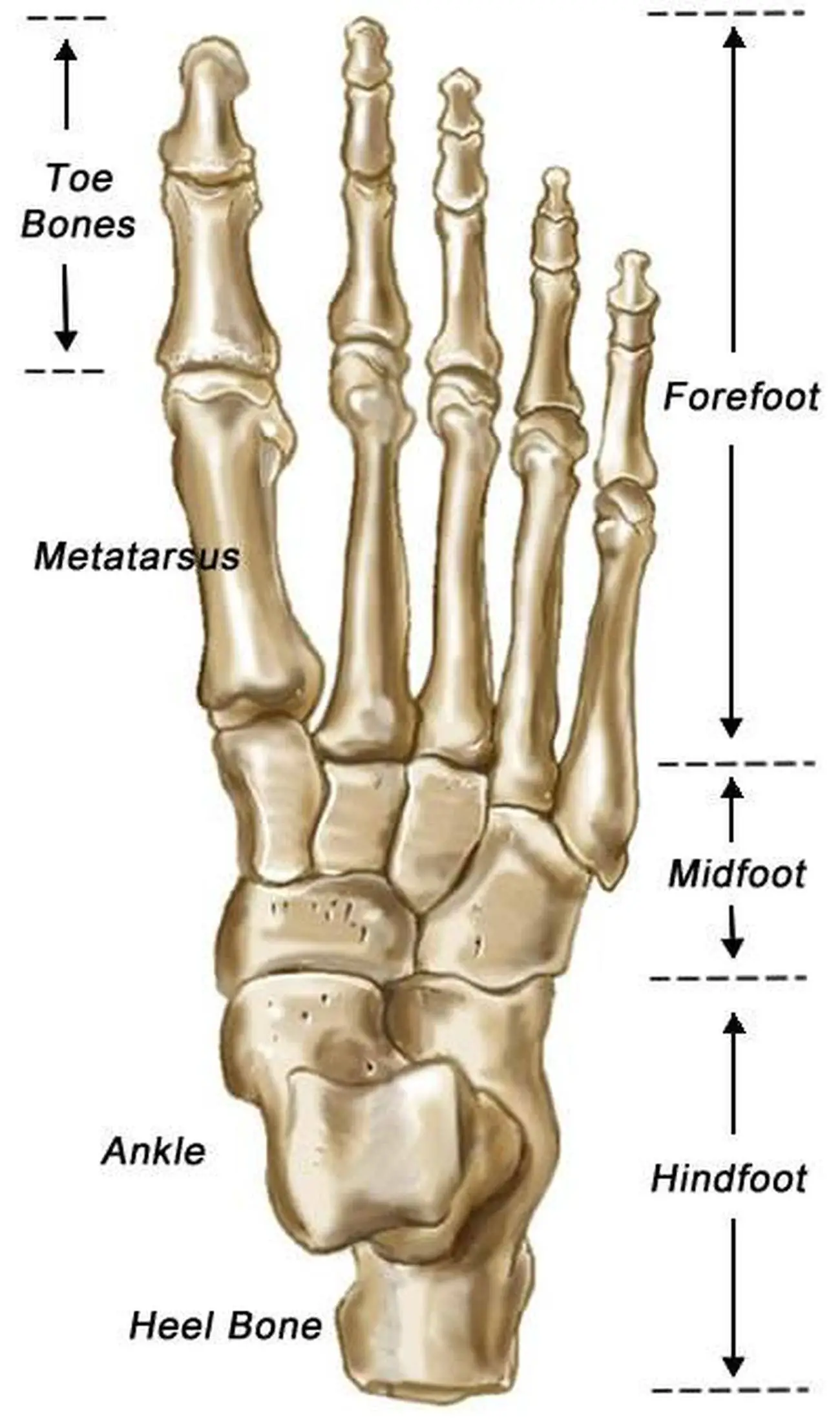
The anatomy of the foot The foot contains a lot of moving parts - 26 bones, 33 joints and over 100 ligaments. The foot is divided into three sections - the forefoot, the midfoot and the hindfoot. The forefoot This consists of five long bones (metatarsal bones) and five shorter bones that form the base of the toes (phalanges).
Calcaneus bone anatomy, function, calcaneus pain & calcaneus fracture
Browse 19,300+ foot anatomy stock photos and images available, or search for foot anatomy vector to find more great stock photos and pictures. Human foot anatomy cutaway representation, clipping path included. "Human foot anatomy cutaway representation, showing skin, veins and arterias, muscles, bones.
Bones of the foot and ankle, medial view with labels App… Flickr
Browse 13,100+ foot bone stock photos and images available, or search for human foot bone or foot bone structure to find more great stock photos and pictures. human foot bone foot bone structure foot bone diagram foot bone vector Sort by: Most popular Human foot bones front and side view anatomy

Ankle and Foot Pain Massage Therapy Connections
The bones of the foot are organized into rows named tarsal bones, metatarsal bones, and phalanges. These make up the toes and broad section of the feet. The other bones of the foot.

Bones In Foot Hottie Fuck
Summary The foot is an intricate part of the body, consisting of 26 bones, 33 joints, 107 ligaments, and 19 muscles. Scientists group the bones of the foot into the phalanges, tarsal.
