Nail Anatomy
Toenails A nail is a horn-like envelope covering the dorsal aspect of the terminal phalanges of fingers and toes in humans, most primates, and a few other mammals. Nails are similar to claws, which are found on numerous other animals. In common usage, the word nail often refers to the nail plate only.

13 best images about Chapter 8 Structure of the Nails on Pinterest Nail strengthening
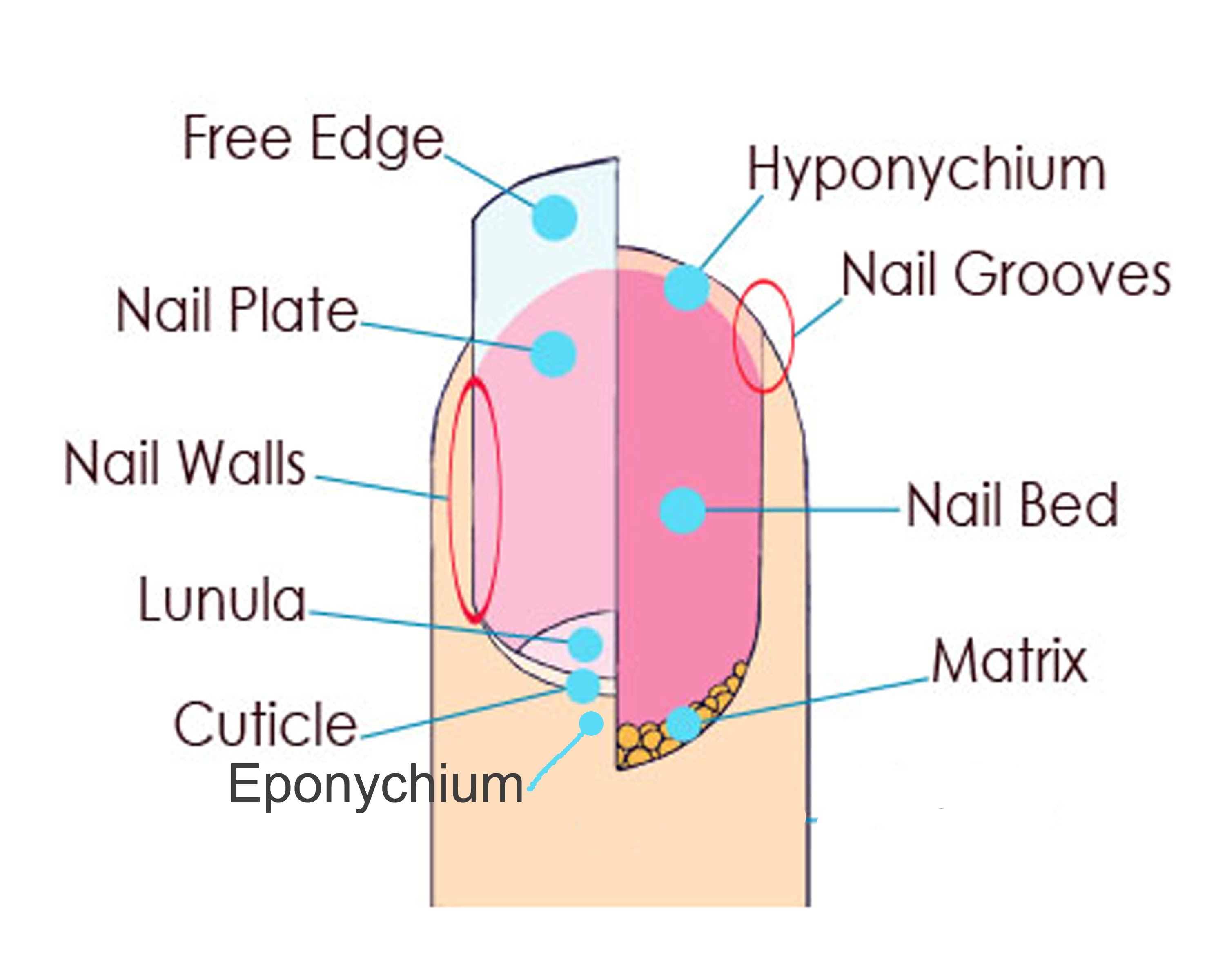
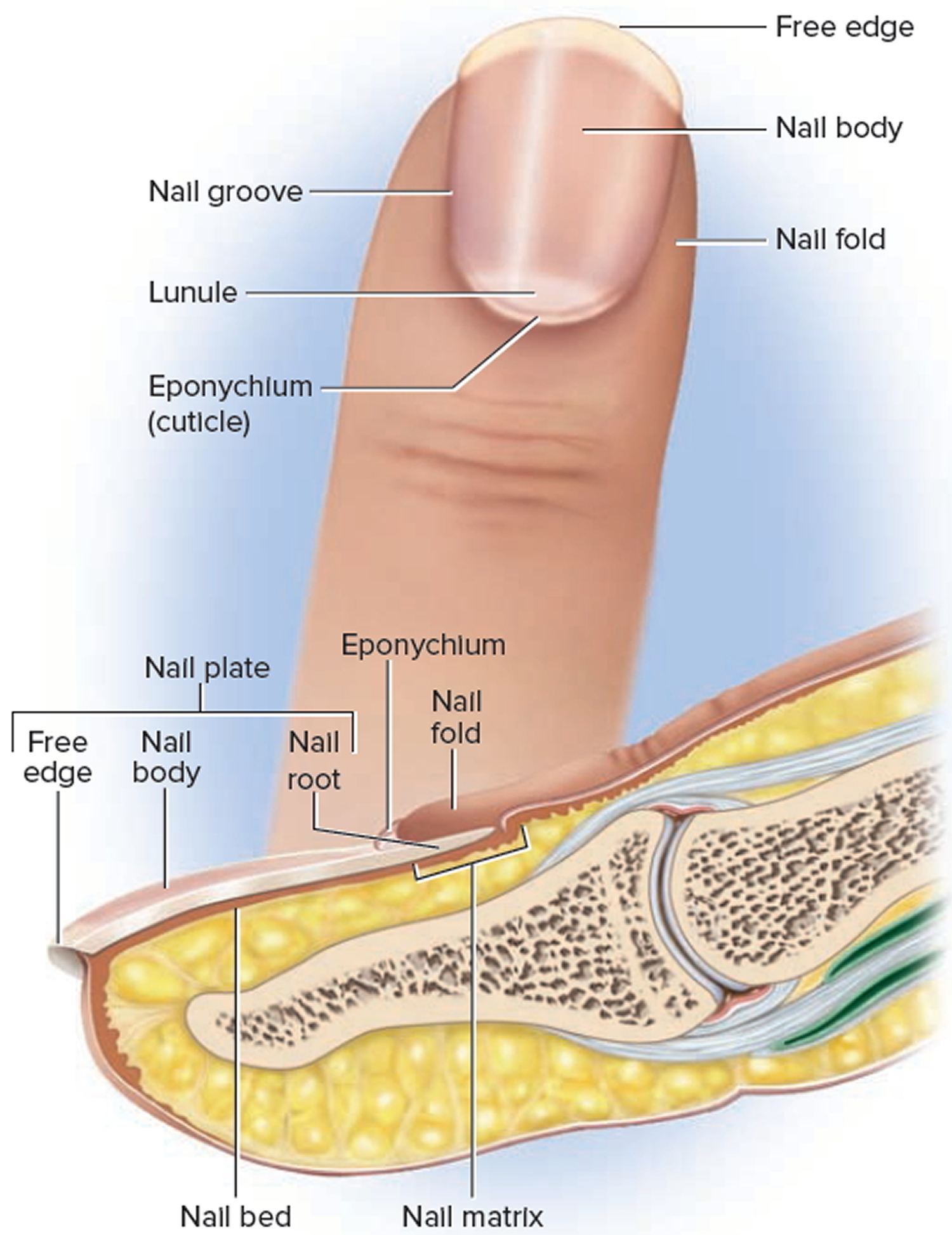
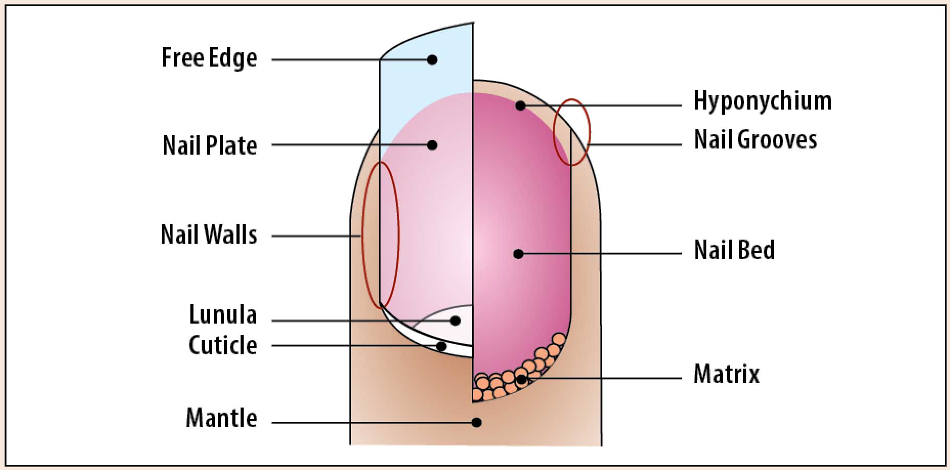
Lunula A bluish-white, opaque area that is visible through the nail plate. This area is the front part of the nail matrix. Sometimes, it's called the "moon." The lunula is the front part of the matrix we can see, or in other words, the visible matrix. Not all fingers have a visible lunula.
Manicure The Ontario Nail Institute
Structure of the nails Created: June 28, 2018; Next update: 2021. Fingernails and toenails are made from skin cells. Structures that are made from skin cells are called skin appendages. Hairs are also skin appendages. The part that we call the nail is technically known as the "nail plate."
Nail Anatomy A Professional Primer on the Parts of the Nail
The nail unit is a complex structure located on the dorsal surface of the fingers and toes. It has two main functions: Protection - protects the digits from trauma Sensation - assists with tactile sensation In this article, we shall look at the anatomy of the nail unit - its component parts and clinical correlations.
The Nail Unit Plate Germinal Matrix Bed TeachMeAnatomy
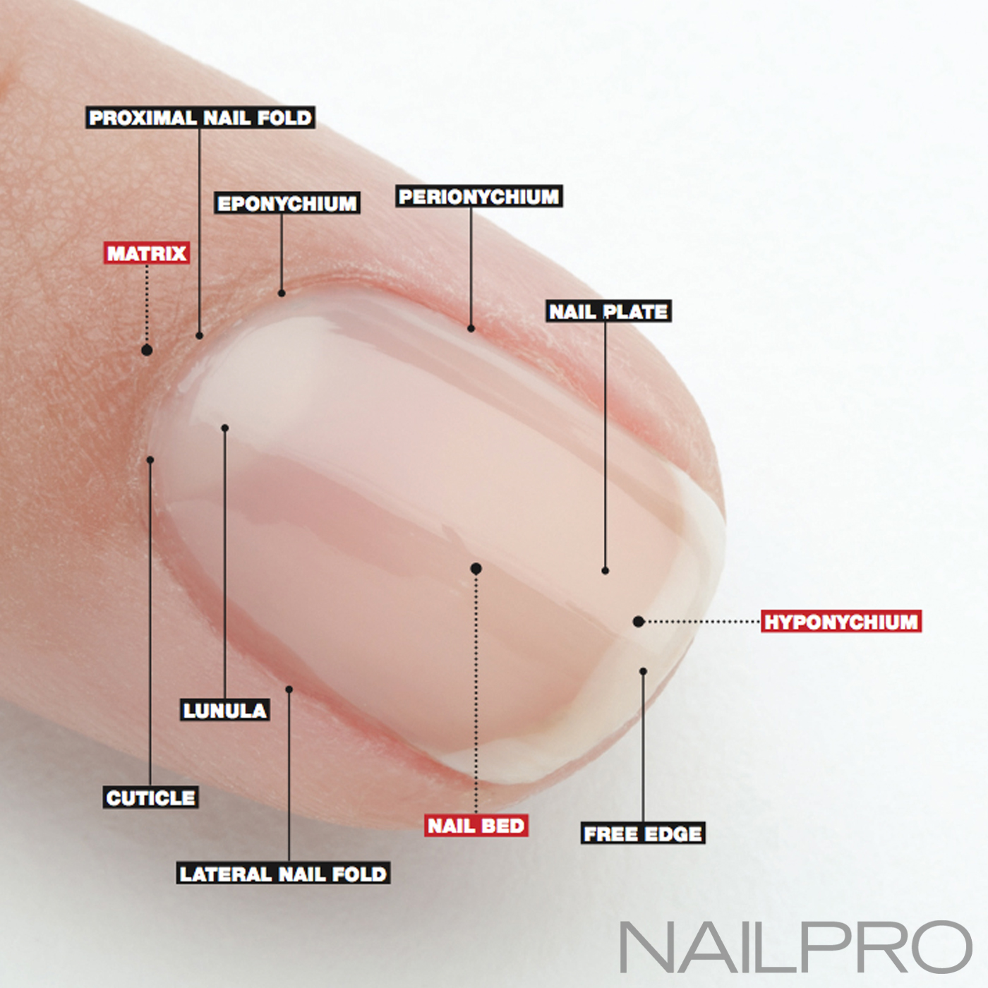
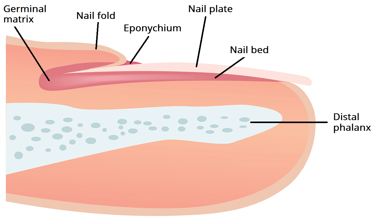
Here, a refresher on the essential parts of the nail, from base to tip and everything in between. Located beneath the skin at the nail's base, the matrix contains nerves and blood and lymph vessels that produce nail cells. The new cells flatten and are pushed forward toward the fingertip resulting in nail growth.
Fingernails Ingrown fingernails Dark Line Fingernail Pain
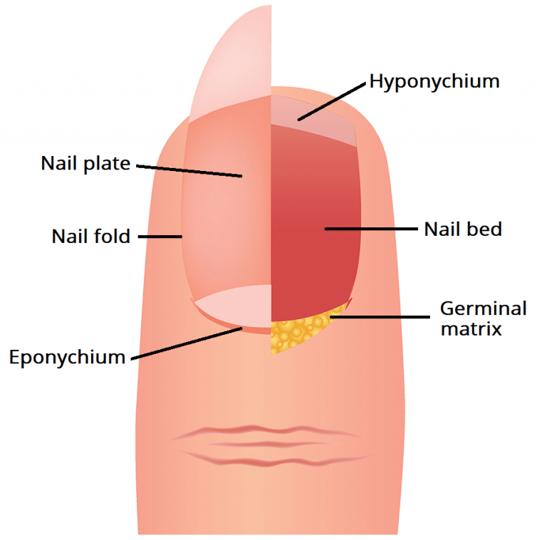
Underlying Structures The nail bed is also referred to as the sterile matrix. It extends from the edge of the nail root, or lunula, to the tissue known as hyponychium . The nail bed contains blood vessels, nerves, and melanocytes that produce melanin.

Nail Structure Diagram Basic Anatomy Of The Nail Unit Dorsal View Download Scientific Diagram
Figure 1. The nail is an accessory structure of the integumentary system. In addition, the nail body forms a back-support for picking up small objects with the fingers. The nail body is composed of densely packed dead keratinocytes. The epidermis in this part of the body has evolved a specialized structure upon which nails can form.

FileHuman nail anatomy.jpg Wikipedia, the free encyclopedia
Nail Plate - This is the thing most people refer to as the fingernail. The largest part of the nail is composed of layers of keratin. The structure is similar to human skin and hair since it is.

Nail Anatomy (and why you should know it)
Nail Anatomy. Figure 10.6.2 The top diagram in this diagram shows the external, visible part of the nail and the cuticle. The bottom diagram shows internal structures in a cross-section of the nail and nail bed. A nail has three main parts: the root, plate, and free margin. Other structures around or under the nail include the nail bed, cuticle.
How To Bill Multiple Nail Evulsions Carr Thert1979
The nail plate consists of close-packed, adherent, interdigitating cells that lack nuclei or organelles. Cells in the plate are very flat, lying with the smallest diameter perpendicular to the plane of the nail plate surface ( Fig. 5.5).There is a progression from the top (dorsal surface) of the plate, where cell borders are straight, to the middle of the plate, where cell borders are much.

Structure of a Nail (CrossSectional View) Diagram Quizlet
A nail consists of: the nail plate, nail folds, nail matrix, nail bed and hyponychium. Nail plate. The nail plate is a rectangular and convex structure embedded within the nail folds. It originates from the nail matrices, found at the base of the nails. The nail plate is completely free distally to the onychodermal band (distal margin of the.
Nail Germinal Matrix Histology nailsr
Diagram Nail bed anatomy Medical conditions Diagnosis Summary What is nail matrix? The nail matrix is the area where your fingernails and toenails start to grow. The matrix creates new.

A longitudinal section showing the structure of a nail. Longitudinal section, Nursing school
Structure. The nail unit includes the nail plate and supporting structures. This section explains the anatomy of the nail unit and histologic findings of each region.. Ink, suture, or an accompanying diagram can facilitate communication of the orientation. Processing a nail biopsy is more challenging than a standard skin biopsy. The specimen.
6.4 Anatomy of the Nails Biology LibreTexts
Overview. The nail organ is an integral component of the digital tip. It is a highly versatile tool that protects the fingertip, contributes to tactile sensation by acting as a counterforce to the fingertip pad, and aids in peripheral thermoregulation via glomus bodies in the nail bed and matrix. [ 1, 2] Because of its form and functionality.

PPT Unit 4 PowerPoint Presentation, free download ID2098133
Structure A. Nail plate; B. lunula; C. root; D. sinus; E. matrix; F. nail bed; G. hyponychium; H. free margin. The nail consists of the nail plate, the nail matrix and the nail bed below it, and the grooves surrounding it. [2] Parts of the nail The nail matrix is the active tissue (or germinal matrix) that generates cells.

diagram of nail anatomy
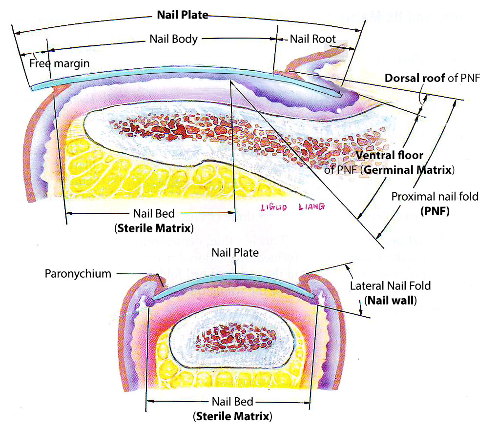
Structure and Function The nail has many soft tissue structures that help support and form the hard-outer nail, known as the nail plate. The attached figure depicts the gross structures described below. Nail Folds The nail folds are soft tissue structures that protect the lateral and proximal edges of the nail plate.
