Ear Anatomy, Facts & Function
human ear, organ of hearing and equilibrium that detects and analyzes sound by transduction (or the conversion of sound waves into electrochemical impulses) and maintains the sense of balance (equilibrium). Understand the science of hearing and how humans and other mammals perceive sound How humans and other mammals perceive sound.
What is Meniere's disease? Hearing Link
1. the externally visible cartilaginous structure of the external ear Click the card to flip 👆 1 / 12 Flashcards Learn Test Match Q-Chat Created by aimige Students also viewed Senses 60 terms anna01017 Preview Psychology Chapter 5: States of Consciousness Teacher 14 terms k_mccroskey Preview Unit 1: Intro to Chemistry Teacher 27 terms Mr__Leppert

15.3 Hearing Anatomy & Physiology
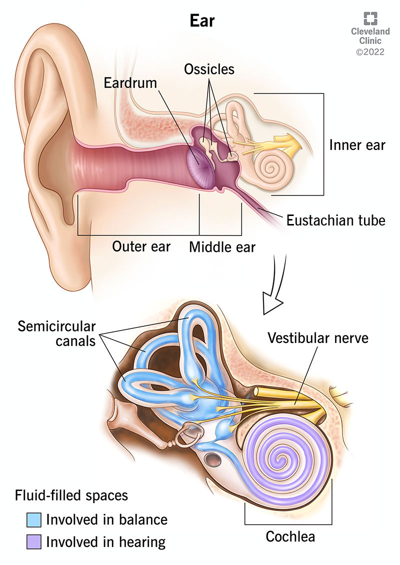
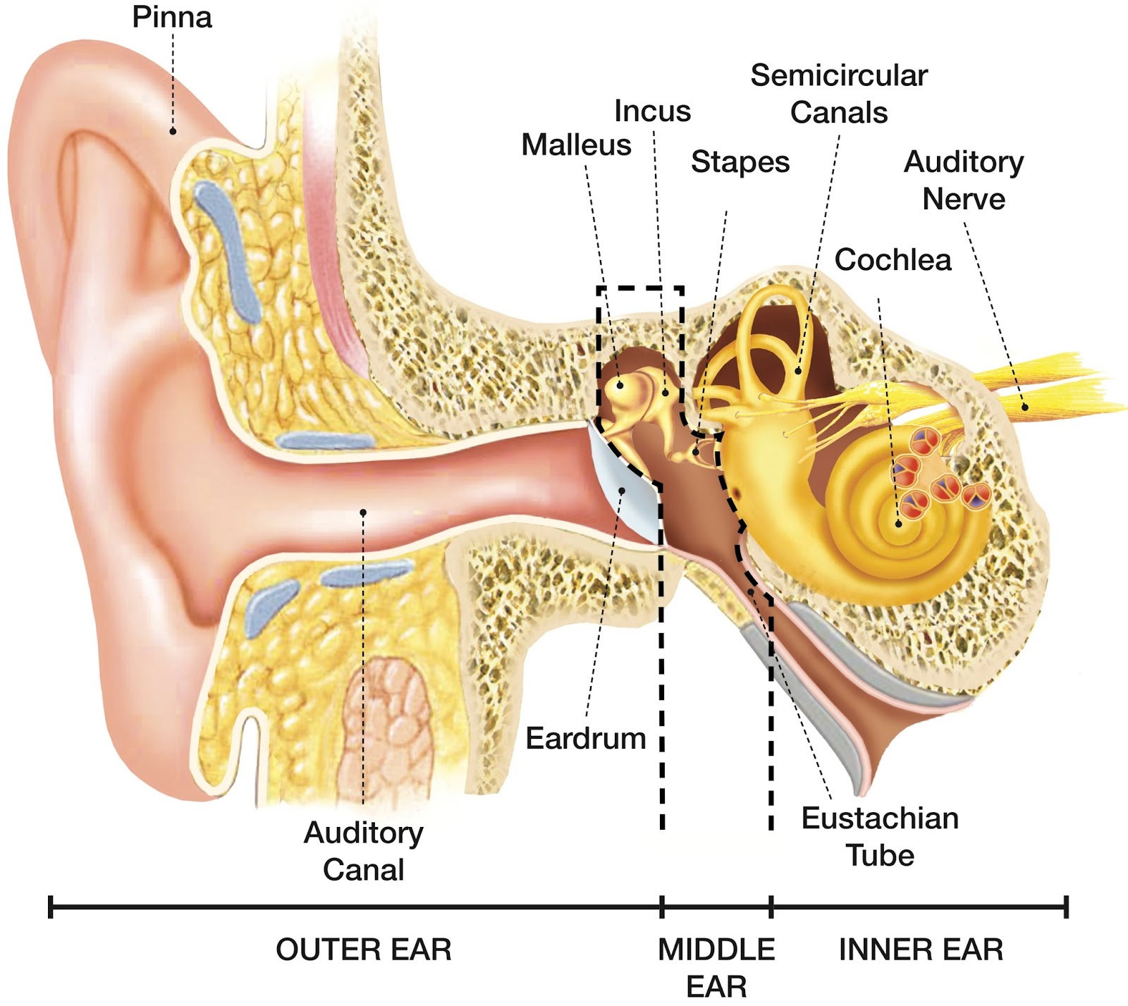
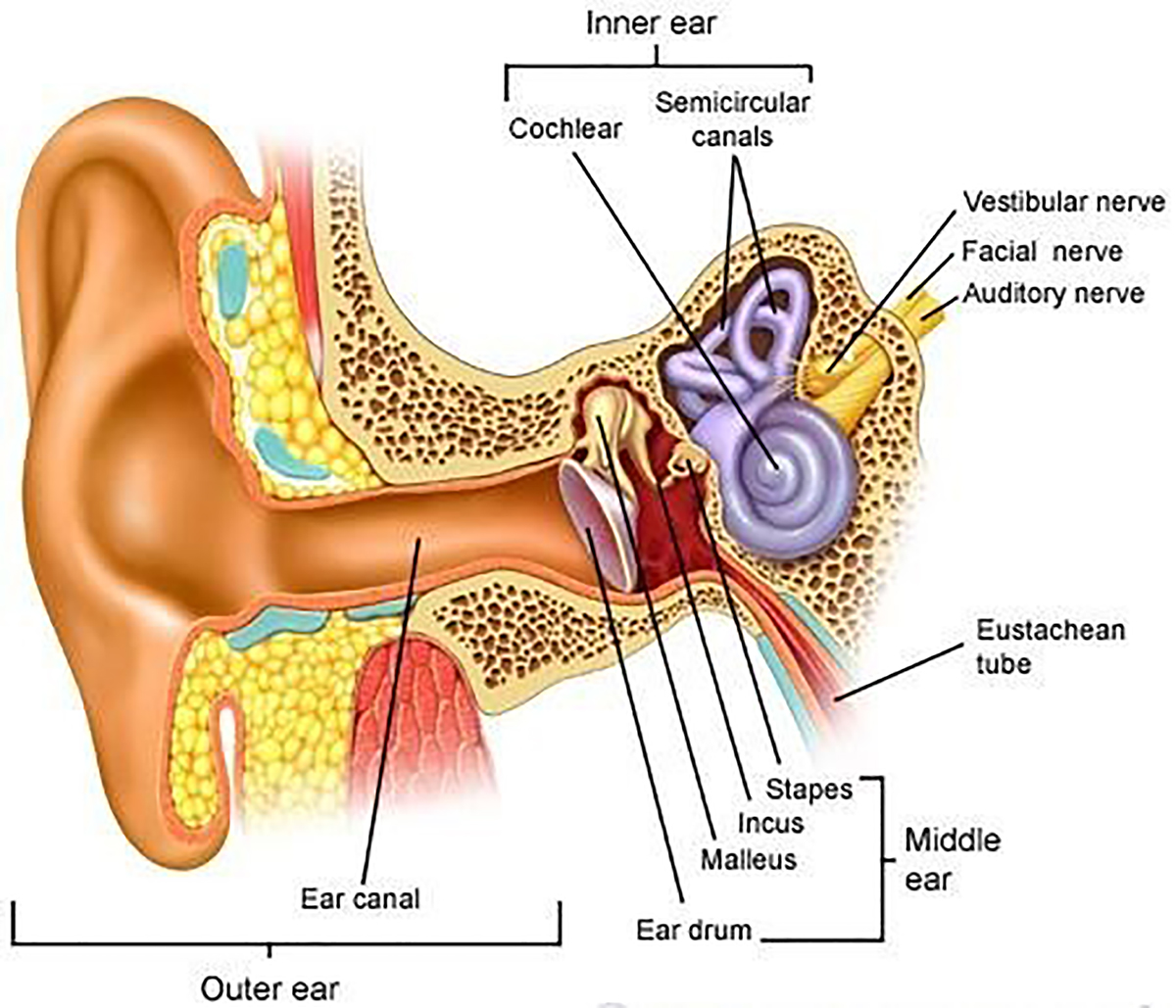
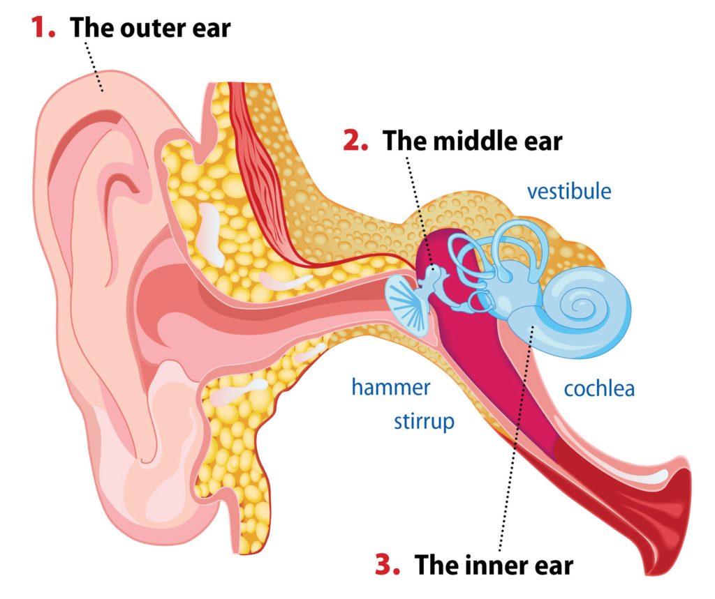
Your outer ear and middle ear are separated by your eardrum, and your inner ear houses the cochlea, vestibular nerve and semicircular canals (fluid-filled spaces involved in balance and hearing). What is the ear? Your ears are organs that detect and analyze sound. Located on each side of your head, they help with hearing and balance. Advertisement
Ear Anatomy Causes of Hearing Loss Hearing Aids Audiology
Middle Ear Anatomy. The middle ear contains most of the small organs responsible for collecting and clarifying external sound waves. It also maintains air pressure balance in the skull through the regulation of the Eustachian tube. The middle ear is most susceptible to ear infections. It can also develop otitis media, which is a category of ear.

Afbeeldingsresultaat voor middle ear anatomy Ear anatomy, Middle ear
The middle ear is the portion of the ear medial to the eardrum, and distal to the oval window of the cochlea (of the inner ear).. The mammalian middle ear contains three ossicles (malleus, incus, and stapes), which transfer the vibrations of the eardrum into waves in the fluid and membranes of the inner ear.The hollow space of the middle ear is also known as the tympanic cavity and is.
How The Ear Works Step by Step Brief Explanation
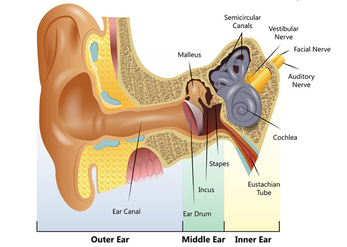
The ear is structurally divided into three parts: the outer (external), middle and inner ear. The middle ear is an air-filled pressurized space within the petrous portion of the temporal bone, extending from the tympanic membrane (eardrum) to the lateral wall of the inner ear.
SPEECH LANGUAGE PATHOLOGY & AUDIOLOGY HEARING DISORDERS OF THE OUTER EAR
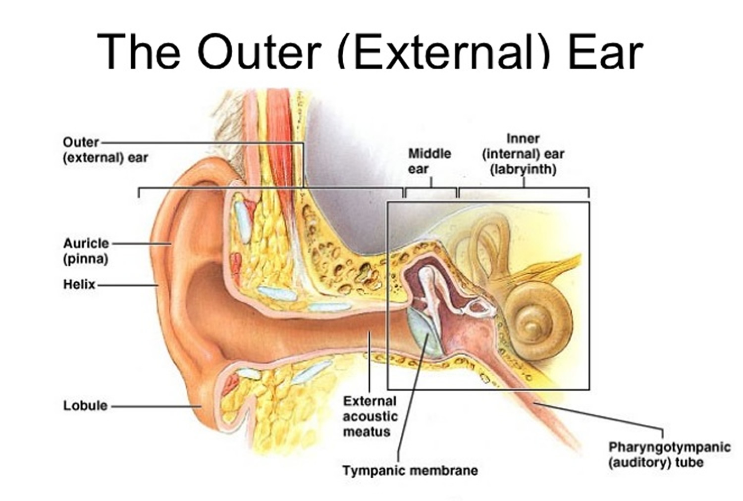
Middle ear Internal ear This mixture of bones, nerves, vessels, membranes, and muscles that make up the ear will be described in this article. Contents External ear Auricle External acoustic meatus Tympanic membrane Muscles of the external ear Vasculature of the external ear Innervation of the external ear Middle ear Tympanic cavity
4.3 Hearing Introduction to Psychology
The ear drum is a transparent membrane which is super sensitive to the vibrations of the ear. So as the air vibrates even the ear drum starts vibrating. Just like the skin of a drum. And as you can, the ear drum also separates the outer ear from the middle ear. This brings us to the middle ear.
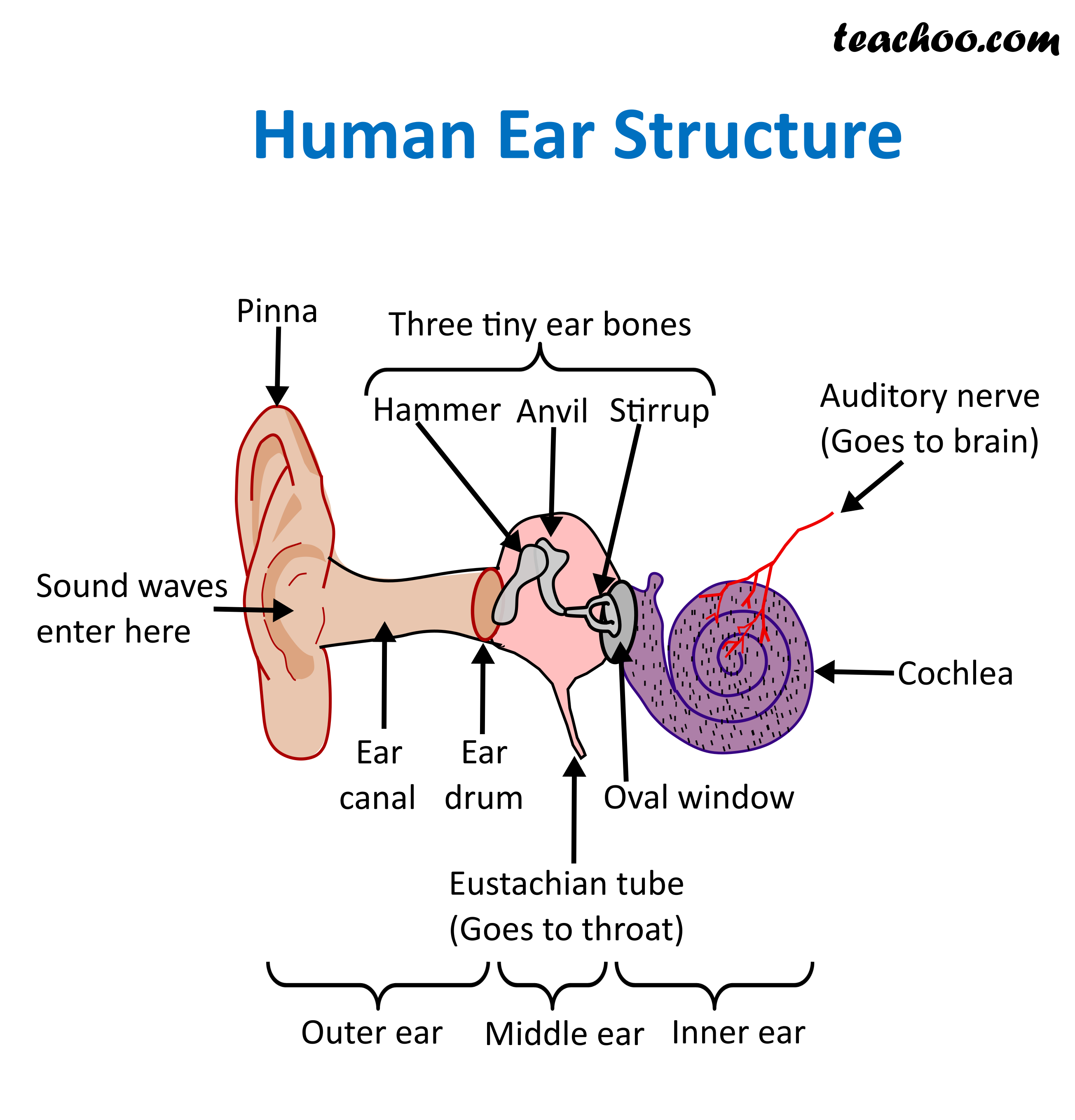
Structure and Function of Human Ear with Diagram Teachoo
Middle ear. Also known as the tympanic cavity, the middle ear is an air-filled, membrane-lined space located between the ear canal and the Eustachian tube, cochlea, and auditory nerve. The eardrum.
earanatomylg Pain Relief Chiropractic
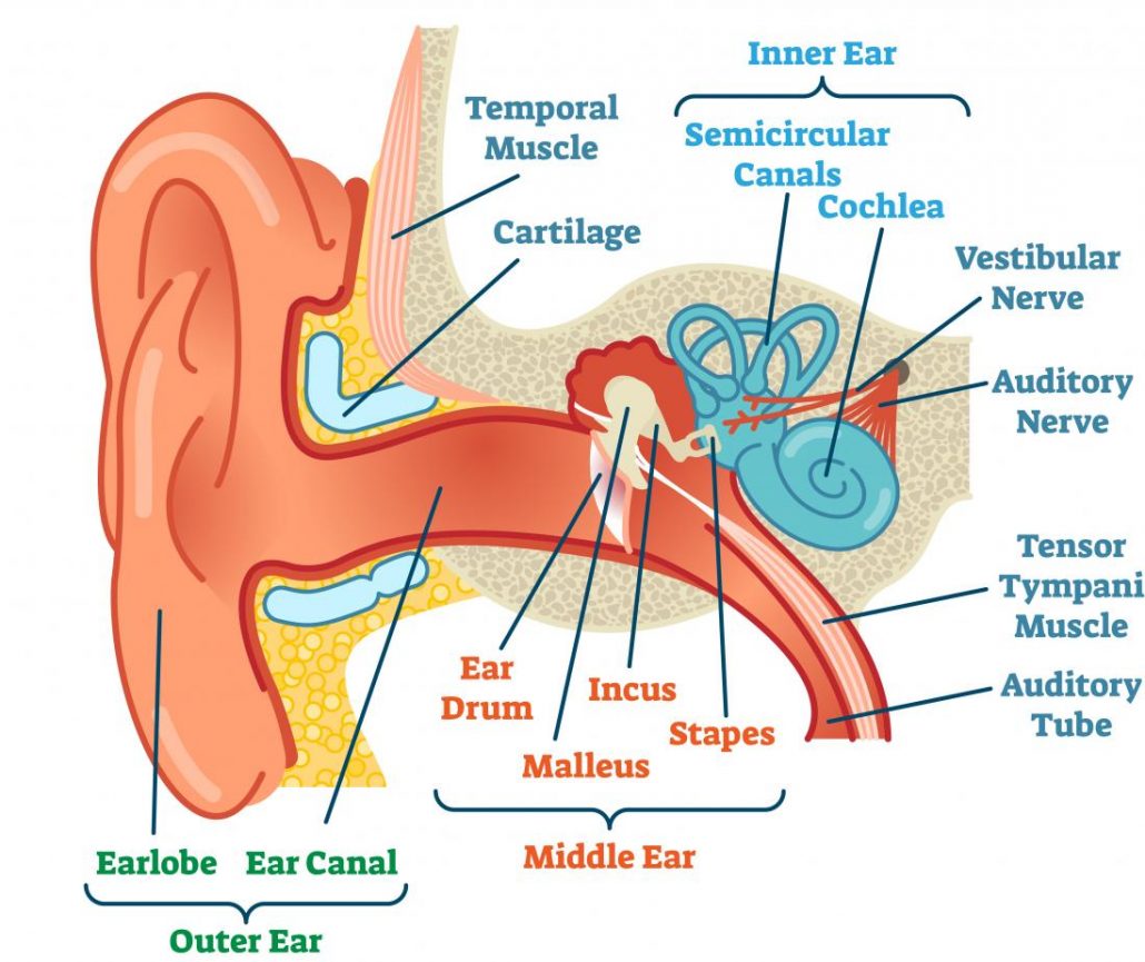
Now, while the inner ear also plays a role in balance, the main role of the external and middle ear is to transfer and amplify sound to the inner ear with the help of the three smallest bones in the body: the auditory ossicles. Let's start with the external ear, which is by far the most common anatomical spot to hang earrings from.
What is conductive hearing loss? Blog of Kiversal
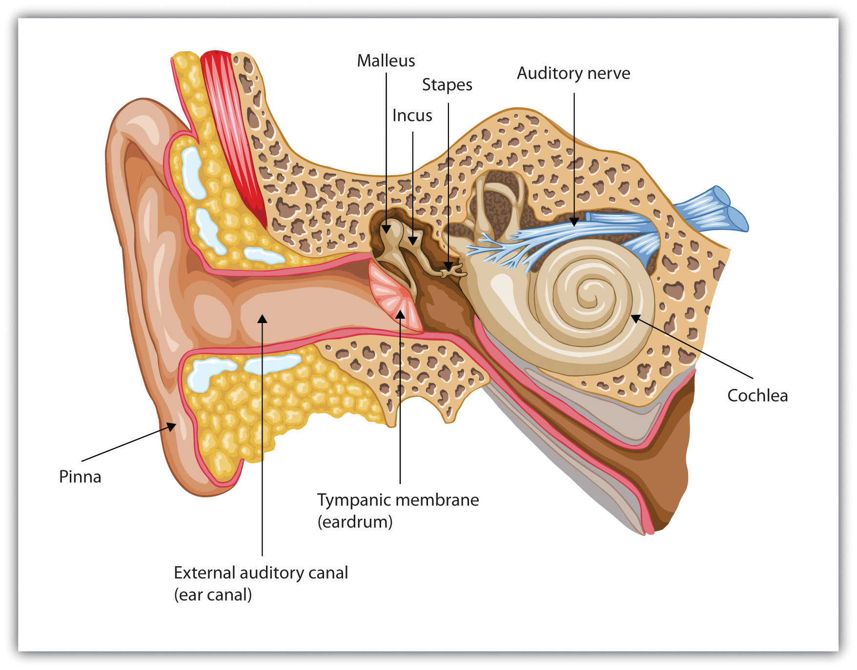
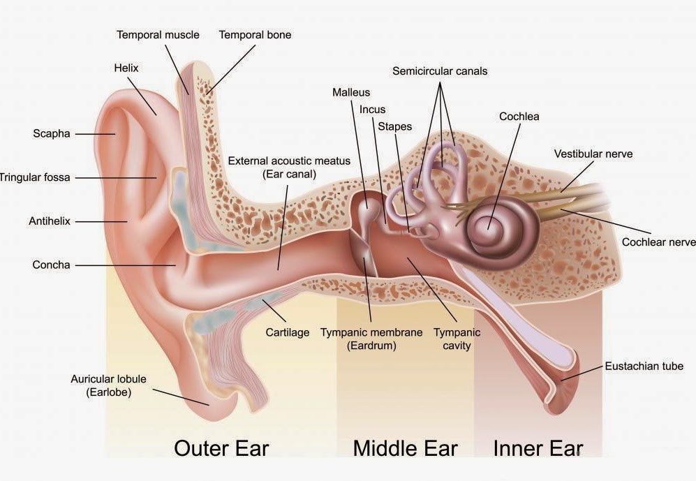
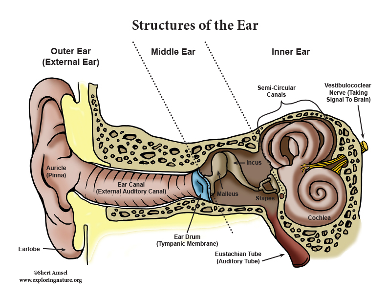
Description: Structures of the Ear. The external ear contains the auricle, ear canal, and tympanic membrane. The middle ear contains the ossicles and is connected to the pharynx by the Eustachian tube. The inner ear contains the cochlea and vestibule, which are responsible for audition and equilibrium, respectively. English labels.

Anatomy Of Ear Labeled How We Perceive Sound Davidson Hearing Aid
It is divided into two parts: Tympanic cavity (adjacent to the tympanic membrane) Epitympanic recess/attic (space superior to the tympanic cavity) Medial (Labyrinthine) Wall The medial (labyrinthine wall) is formed by the lateral aspect of the inner ear.

Human ear anatomy. Ears inner structure, organ of hearing ve (1000410
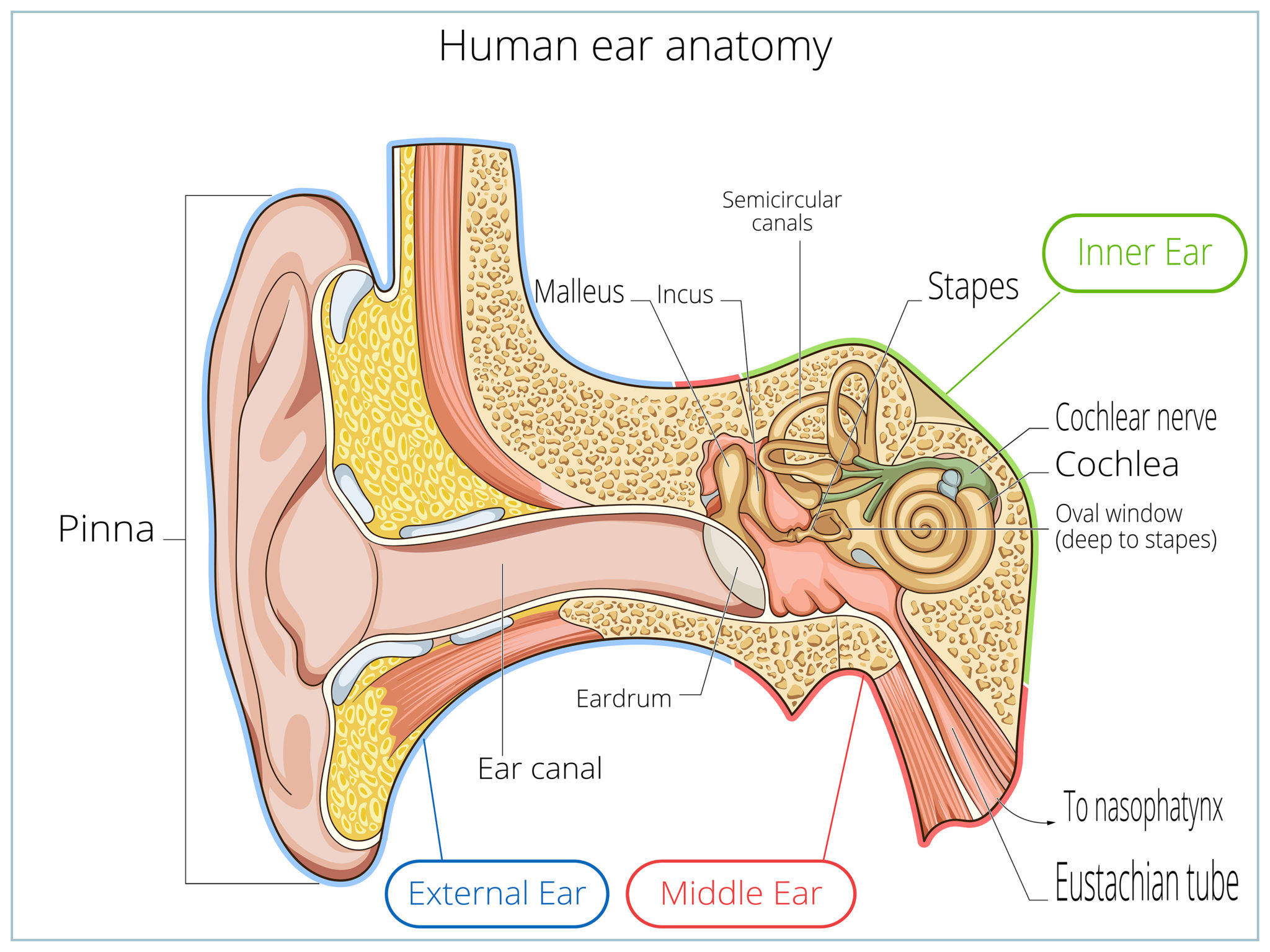
The middle ear's auditory ossicles are a chain-like arrangement of three tiny bones that extend from the tympanic membrane to the oval window. The three auditory ossicles are: Malleus or hammer - It has a head, a neck, and a hand. The manubrium or hand is joined to the tympanic membrane. The neck stretches from the hand to the head.
Outer Ear Anatomy Outer Ear Infection & Pain Causes & Treatment
Parts of the Middle Ear The middle ear is an air-filled cavity that sits between the tympanic membrane [3] and the inner ear. The middle ear also consists of three tiny bones called ossicles [4], the round window [5], the oval window [6], and the Eustachian tube [7] . Ossicles and Their Function Malleus (commonly known as the hammer)
The ear structure and functions Blog of Kiversal
The structures in the ear are divided into three categories: the outer ear, the middle ear, and the inner ear. Let's go through them one by one and learn more about ear anatomy. Outer ear. The outermost part of the ear is the auricle, also known as the external ear or the pinna. Made of cartilage covered in thin skin, the auricle amplifies.
Hearing and the Structure of the Ear
That's why labeling the ear is an effective way to begin your revision. It helps you to memorize the names and their locations, which in turn will aid you to remember their functions. Below, you can download both the blank ear diagram to make some notes, and then try labeling the ear using the unlabeled ear diagram. Good luck!
